Review
. 2021 May 29;22(11):5843.
doi: 10.3390/ijms22115843.

Erythrocytes: Central Actors in Multiple Scenes of Atherosclerosis

Affiliations
Review

Erythrocytes: Central Actors in Multiple Scenes of Atherosclerosis

Chloé Turpin et al. Int J Mol Sci. .

Abstract

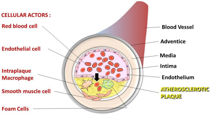
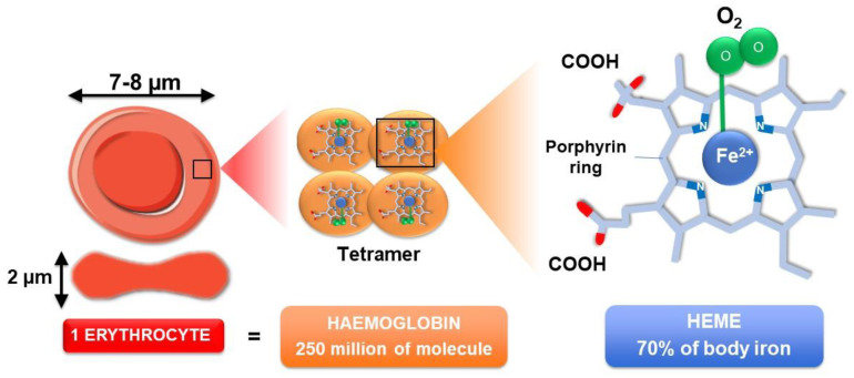
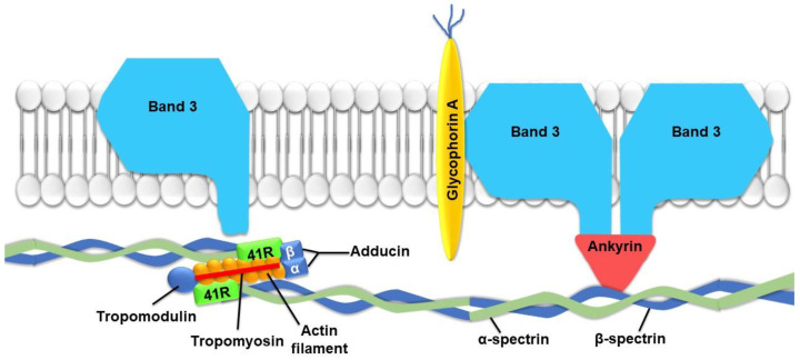
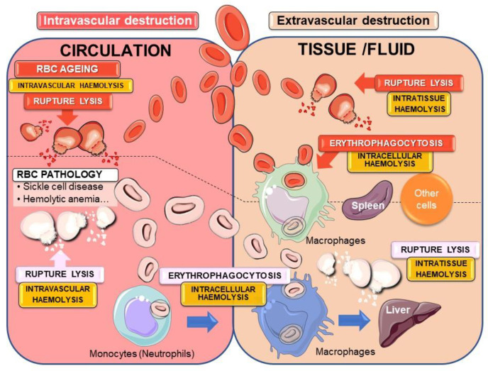
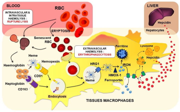
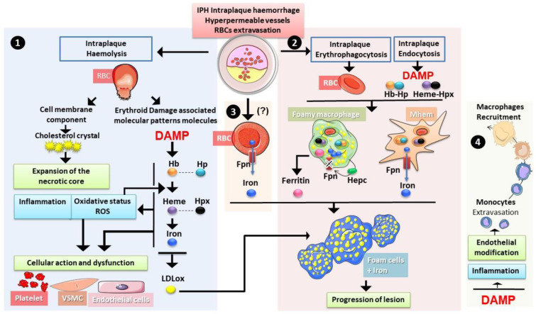
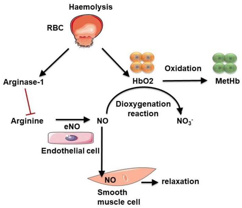
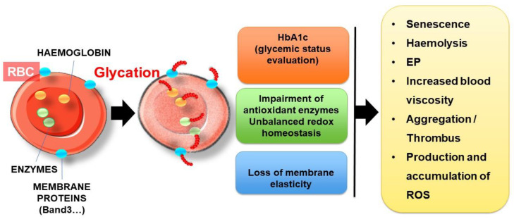
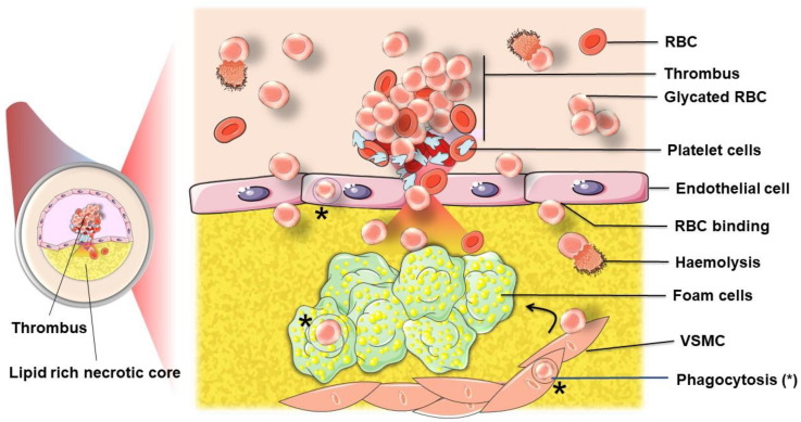
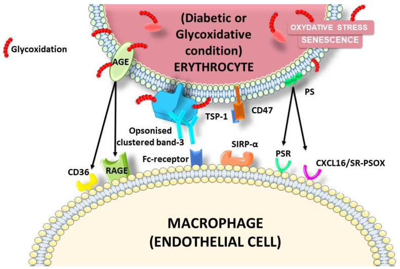
The development and progression of atherosclerosis (ATH) involves lipid accumulation, oxidative stress and both vascular and blood cell dysfunction. Erythrocytes, the main circulating cells in the body, exert determinant roles in the gas transport between tissues. Erythrocytes have long been considered as simple bystanders in cardiovascular diseases, including ATH. This review highlights recent knowledge concerning the role of erythrocytes being more than just passive gas carriers, as potent contributors to atherosclerotic plaque progression. Erythrocyte physiology and ATH pathology is first described. Then, a specific chapter delineates the numerous links between erythrocytes and atherogenesis. In particular, we discuss the impact of extravasated erythrocytes in plaque iron homeostasis with potential pathological consequences. Hyperglycaemia is recognised as a significant aggravating contributor to the development of ATH. Then, a special focus is made on glycoxidative modifications of erythrocytes and their role in ATH. This chapter includes recent data proposing glycoxidised erythrocytes as putative contributors to enhanced atherothrombosis in diabetic patients.

Keywords: atherosclerosis; eryptosis; erythrocytes; erythrophagocytosis; glycation; haemoglobin; heme; iron; oxidative stress.


Conflict of interest statement

The authors declare no conflict of interest.

Figures

Figure 1
Figure 1
Cellular actors present in the atheroma plaque. Dysfunction of endothelial cells allows lipoprotein and erythrocyte infiltration in the sub-endothelial space (the arrow indicates this infiltration). Intraplaque macrophages that engulf oxidised LDL and infiltrated erythrocytes differentiate into foam cells initiating the atheroma formation. Enhanced inflammatory processes in atheroma cause smooth muscle cell migration from the media to the intima, towards the necrotic core formed by dead foam cells.
Figure 2
Figure 2
Erythrocyte structure and content. One single erythrocyte contains about 250 million haemoglobin molecules. Haemoglobin, the protein responsible for oxygen transport, is a tetramer composed of 4 globin chains. Each globin chain contains one heme molecule responsible for the binding of one oxygen molecule (O2). Heme consists of a porphyrin ring centred by one atom of iron (Fe2+). Oxygen strongly binds to this iron atom. Almost two-thirds of the body iron (about 2.5 to 36 g) is localised in haemoglobin of circulating erythrocytes.
Figure 3
Figure 3
Schematic representation of important proteins involved in erythrocyte membrane flexibility. Spectrin molecules, responsible for stability and elasticity, are attached to the cell membrane by ankyrin. 41R protein establishes a link between actin and spectrin filaments.
Figure 4
Figure 4
Physiological and pathological intra- and extravascular destruction of erythrocytes. The fate of senescent (or eryptotic) red blood cells (RBCs) can undergo different pathways for cellular destruction. Rupture lysis into the blood circulation (intravascular haemolysis) or into tissues and fluids (intratissue haemolysis) in the case of RBC intravasation can occur with the liberation of hemolytic by-products. To avoid such release of harmful components, the major mechanism of senescent erythrocyte removal passes through the process of erythrophagocytosis by macrophages (intracellular haemolysis) mainly in the spleen. In many erythrocyte pathologies, the process of eryptosis is accelerated leading to massive elimination of erythrocytes both by rupture lysis and erythrophagocytosis into the circulation or inside tissues/fluid as well. Pathological erythrocytes can be recognised and phagocytosed by circulating monocytes that could migrate to tissues such as the liver.
Figure 5
Figure 5
Erythrocyte haemolysis and heme iron recycling. Modifications (oxidative stress, glycation; see chapter IV) and/or eryptosis can trigger the lysis of circulating or extravasated erythrocytes in some tissues. After such intravascular or intratissue haemolysis, haptoglobin and hemopexin bind to haemoglobin and heme, respectively. Complexes are recognised by macrophages and endocytosed via their specific receptors CD163 for haemoglobin/haptoglobin complexes and CD91 for heme/hemopexin complexes. In the case of EP of senescent or eryptotic erythrocytes (intracellular haemolysis), specific recognition of old erythrocytes is performed by tissue macrophages, and a phagolysosome containing the old erythrocyte is formed. Both CD163- and CD91-mediated endocytosis and EP lead to the formation of a vacuole containing heme. Heme is then transported to the cytosol via HRG1 and degraded by HMOX1 (Heme oxygenase 1) to produce iron. According to the body needs, such iron is either stored in ferritin or transported outside the cells via the transporter ferroportin. Hepcidin, a small but powerful regulatory peptide of iron homeostasis, is mainly produced by hepatocytes and to a lesser extent by immune cells such as macrophages. Hepcidin binds to ferroportin inducing its internalization and degradation. Such interaction is primordial for regulating macrophage iron levels as well as systemic iron homeostasis.
Figure 6
Figure 6
Erythrocytes in intraplaque haemorrhage. During intraplaque haemolysis 1. and rupture lysis, RBCs liberate both cholesterol and erythroid damage-associated signalling molecule patterns ((DAMPs) haemoglobin Hb, heme and iron), both contributing to the expansion of the necrotic core and to the increase in oxidative stress and inflammation. Such changes could have an impact on the function of endothelial cells, vascular smooth muscle cells (VSMC) and platelets. Haemoglobin and iron can promote the oxidation of LDL particles accelerating the foam cell differentiation. Erythrophagocytosis as well as endocytosis of heme/hemopexin and haemoglobin/haptoglobin complexes 2. also occurs in the plaque with an important role of macrophages. The haemorrhage-associated macrophages (Mhem) are prone to recycling iron from both erythrophagocytosis and heme containing-complexes endocytosis, whereas foamy macrophages (Mox) tend to accumulate iron with lipids. Autocrine expression of hepcidin (Hepc) seems to play a role in iron retention. Ferritin secretion by these cells has been reported. Another source of iron could exist directly from erythrocytes via the expression of ferroportin (Fpn) 3. Both iron and ferritin present in the plaque with oxidised low-density lipoprotein (oxLDL) could trigger the formation of foam cells, promoting the progression of the lesion and the instability of the plaque. In addition, 4. erythroid DAMPs were shown to increase expression of adhesion molecules by endothelial cells and to have proinflammatory effects. Both phenomena contribute to the recruitment of more monocytes/macrophages in the plaque.
Figure 7
Figure 7
Influence of haemolysis on nitric oxide (NO) bioavailability. Intravascular haemolysis induces arginase-1 and haemoglobin release. Arginase-1 produces ornithine from arginine, the required substrate for NO production. Oxygenated haemoglobin (HbO2) can scavenge NO by a dioxygenation reaction, producing inert nitrate and methemoglobin (MetHb). These two reactions decrease NO bioavailability.
Figure 8
Figure 8
Deleterious effects of erythrocyte glycation. Hyperglycaemia causes glycation of many RBC components, such as haemoglobin, antioxidant enzymes and cell surface membrane proteins. This phenomenon induces unbalanced redox homeostasis with the loss of membrane elasticity. As a consequence, erythrocyte senescence, haemolysis, EP, erythrocyte aggregation and ROS production are enhanced.
Figure 9
Figure 9
Participation of RBCs to atherothrombosis. Following plaque rupture, RBCs can further infiltrate the plaque where they are either phagocytosed (*) by endothelial cells, macrophages and vascular smooth muscle cells (VSMC), or lysed. Erythrocytes, by their adhesive and aggregation properties, exacerbated when glycated, also contribute, with platelets, to the thrombus formation. When glycated or aged, phosphatidylserine exposing erythrocytes can further interact with activated platelets [182]. This process participates in the occlusive phenomenon.
Figure 10
Figure 10
Glycation induces senescence and haemolysis of erythrocytes. Erythrocyte smears stained with May-Grünwald Giemsa stain for (A) normal erythrocytes. There is no porous structure in haemoglobin repartition in non-glycated erythrocytes. (B) Glycated erythrocytes incubated with 50 mM of glucose for 5 days at 37 °C display different morphologies. Arrows 1 feature erythrocytes with non-homogeneous structure and repartition of haemoglobin with presence of vacuoles. Arrows 2 feature erythrocytes with more condensed haemoglobin and a peripheral clear area. Arrows 3 feature erythrocyte ghosts corresponding to senescent RBCs after the release of haemoglobin following haemolysis. Bars indicate 10 μm.
Figure 11
Figure 11
Ligands and receptors involved in glycated erythrocyte interactions with phagocytic cells. Several changes in the erythrocyte membrane are induced by glycation, such as PS exposure, CD47 shift, AGE formation and band.3 clustering. All these changes can be considered as ligands for the recognition and clearance of glycated/damaged erythrocytes.

References

    1. Benjamin E.J., Muntner P., Alonso A., Bittencourt M.S., Callaway C.W., Carson A.P., Chamberlain A.M., Chang A.R., Cheng S., Das S.R., et al. Heart Disease and Stroke Statistics-2019 Update: A Report From the American Heart Association. Circulation. 2019;139:e56–e528. doi: 10.1161/CIR.0000000000000659. - DOI - PubMed
    1. Palasubramaniam J., Wang X., Peter K. Myocardial Infarction-From Atherosclerosis to Thrombosis. Arterioscler. Thromb. Vasc. Biol. 2019;39:e176–e185. - PubMed
    1. Michel J.B., Martin-Ventura J.L. Red Blood Cells and Hemoglobin in Human Atherosclerosis and Related Arterial Diseases. Int. J. Mol. Sci. 2020;21:6756. - PMC - PubMed
    1. Pasterkamp G., Virmani R. The erythrocyte: A new player in atheromatous core formation. Heart. 2002;88:115–116. doi: 10.1136/heart.88.2.115. - DOI - PMC - PubMed
    1. Arias C.F. How do red blood cells know when to die? R. Soc. Open Sci. 2017;4:160850. doi: 10.1098/rsos.160850. - DOI - PMC - PubMed