Review
. 2021 May 31;22(11):5887.
doi: 10.3390/ijms22115887.

Glucose Metabolic Dysfunction in Neurodegenerative Diseases-New Mechanistic Insights and the Potential of Hypoxia as a Prospective Therapy Targeting Metabolic Reprogramming

Affiliations
Review

Glucose Metabolic Dysfunction in Neurodegenerative Diseases-New Mechanistic Insights and the Potential of Hypoxia as a Prospective Therapy Targeting Metabolic Reprogramming

Rongrong Han et al. Int J Mol Sci. .

Abstract

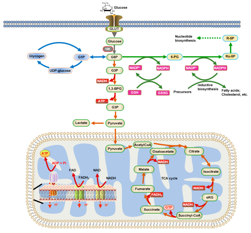
Glucose is the main circulating energy substrate for the adult brain. Owing to the high energy demand of nerve cells, glucose is actively oxidized to produce ATP and has a synergistic effect with mitochondria in metabolic pathways. The dysfunction of glucose metabolism inevitably disturbs the normal functioning of neurons, which is widely observed in neurodegenerative disease. Understanding the mechanisms of metabolic adaptation during disease progression has become a major focus of research, and interventions in these processes may relieve the neurons from degenerative stress. In this review, we highlight evidence of mitochondrial dysfunction, decreased glucose uptake, and diminished glucose metabolism in different neurodegeneration models such as Alzheimer's disease (AD), Parkinson's disease (PD), amyotrophic lateral sclerosis (ALS), and Huntington's disease (HD). We also discuss how hypoxia, a metabolic reprogramming strategy linked to glucose metabolism in tumor cells and normal brain cells, and summarize the evidence for hypoxia as a putative therapy for general neurodegenerative disease.

Keywords: brain energy metabolism; glucose; hypoxia; metabolic reprogramming; neurodegenerative disease.


Conflict of interest statement

The authors declare no conflict of interest.

Figures

Figure 1
Figure 1
Glucose metabolism and energy homeostasis in the brain. Glucose enters the cell through GLUT and is converted to G6P by HK. Then, G6P can be processed by different metabolic pathways: (1) Glycolysis (shown by red arrows), which leads to lactic acid production or the tricarboxylic acid (TCA) cycle. NADH and FADH2 are subsequently re-oxidized in ETC to produce ATP. (2) The pentose phosphate pathway (PPP) (shown by green arrows), which metabolizes G6P and generates NADPH. NADPH is then used for oxidative stress defense and biosynthetic reactions. (3) Glycogenesis (shown by blue arrows). Abbreviations are as follows: GLUT: glucose transporters; HK: Hexokinase; G6P: glucose-6-phosphate; G1P: glucose-1-phosphate; 6-PG: 6-phosphogluconate; G3P: glyceraldehyde-3-phosphate; 1,3-BPG: 1,3-bisphosphoglycerate; Ru-5P: ribulose-5-phosphate; R-5P: ribose-5-phosphate.
Figure 2
Figure 2
The crosstalk of metabolic dysfunction and neurodegenerative disease. Accumulating evidence has suggested the presence of a strong correlation between metabolic dysregulation and neurodegenerative disorders, such as AD, PD, ALS, and HD.
Figure 3
Figure 3
Glucose metabolic reprogramming induced by hypoxia. In normoxia conditions, HIF is constitutively made and hydroxylated by prolyl-hydroxylase (PHD) enzymes; the hydroxylated form is recognized by ubiquitin ligase, Von Hippel-Lindau (VHL), and targeted for proteasomal degradation. In response to the environmental hypoxia, the PHD reaction does not take place, allowing HIF stabilization and translocation to the nucleus. HIF binds with other transcriptional factors to enhance the transcription of genes encoding the enzymes involved in glycolysis.

Similar articles

Cited by

References

    1. Rossi S., Zanier E.R., Mauri I., Columbo A., Stocchetti N. Brain temperature, body core temperature, and intracranial pressure in acute cerebral damage. J. Neurol. Neurosurg. Psychiatry. 2001;71:448–454. doi: 10.1136/jnnp.71.4.448. - DOI - PMC - PubMed
    1. Mergenthaler P., Lindauer U., Dienel G.A., Meisel A. Sugar for the brain: The role of glucose in physiological and pathological brain function. Trends Neurosci. 2013;36:587–597. doi: 10.1016/j.tins.2013.07.001. - DOI - PMC - PubMed
    1. Jain I.H., Zazzeron L., Goli R., Alexa K., Schatzman-Bone S., Dhillon H., Goldberger O., Peng J., Shalem O., Sanjana N.E., et al. Hypoxia as a therapy for mitochondrial disease. Science. 2016;352:54–61. doi: 10.1126/science.aad9642. - DOI - PMC - PubMed
    1. Ashrafi G., Wu Z., Farrell R.J., Ryan T.A. GLUT4 Mobilization Supports Energetic Demands of Active Synapses. Neuron. 2017;93:606–615.e3. doi: 10.1016/j.neuron.2016.12.020. - DOI - PMC - PubMed
    1. Simpson I.A., Carruthers A., Vannucci S.J. Supply and demand in cerebral energy metabolism: The role of nutrient transporters. J. Cereb. Blood Flow Metab. 2007;27:1766–1791. doi: 10.1038/sj.jcbfm.9600521. - DOI - PMC - PubMed

MeSH terms