. 2021 May 31;13(6):1034.
doi: 10.3390/v13061034.

Genomic Analysis of a Novel Phage Infecting the Turkey Pathogen Escherichia coli APEC O78 and Its Endolysin Activity

Affiliations

Genomic Analysis of a Novel Phage Infecting the Turkey Pathogen Escherichia coli APEC O78 and Its Endolysin Activity

Sangsang Deng et al. Viruses. .

Abstract

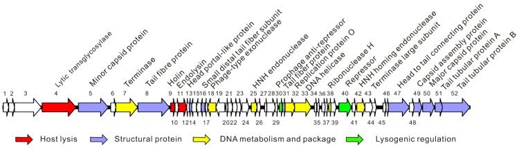
Due to the increasing spread of multidrug-resistant (MDR) bacteria, phage therapy is considered one of the most promising methods for addressing MDR bacteria. Escherichia coli lives symbiotically in the intestines of humans and some animals, and most strains are beneficial in terms of maintaining a healthy digestive tract. However, some E. coli strains can cause serious zoonotic diseases, including diarrhea, pneumonia, urinary tract infections, and hemolytic uremic syndrome. In this study, we characterized a newly isolated Myoviridae phage, vB_EcoM_APEC. The phage vB_EcoM_APEC was able to infect E. coli APEC O78, which is the most common MDR E. coli serotype in turkeys. Additionally, the phage's host range included Klebsiella pneumoniae and other E. coli strains. The genome of phage vB_EcoM_APEC (GenBank accession number MT664721) was 35,832 bp in length, with 52 putative open reading frames (ORFs) and a GC content of 41.3%. The genome of vB_EcoM_APEC exhibited low similarity (79.1% identity and 4.0% coverage) to the genome of Acinetobacter phage vB_AbaM_IME284 (GenBank no. MH853787.1) according to the nucleotide Basic Local Alignment Search Tool (BLASTn). Phylogenetic analysis revealed that vB_EcoM_APEC was a novel phage, and its genome sequence showed low similarity to other available phage genomes. Gene annotation indicated that the protein encoded by orf11 was an endolysin designated as LysO78, which exhibited 64.7% identity (91.0% coverage) with the putative endolysin of Acinetobacter baumannii phage vB_AbaM_B9. The LysO78 protein belongs to glycoside hydrolase family 19, and was described as being a chitinase class I protein. LysO78 is a helical protein with 12 α-helices containing a large domain and a small domain in terms of the predicted three-dimensional structure. The results of site-directed mutagenesis indicated that LysO78 contained the catalytic residues E54 and E64. The purified endolysin exhibited broad-spectrum bacteriolytic activity against Gram-negative strains, including the genera Klebsiella, Salmonella, Shigella, Burkholderia, Yersinia, and Pseudomonas, as well as the species Chitinimonas arctica, E. coli, Ralstonia solanacearum, and A. baumannii. An enzymatic assay showed that LysO78 had highly lytic peptidoglycan hydrolases activity (64,620,000 units/mg) against E. coli APEC O78, and that LysO78 had lytic activity in the temperature range of 4-85 °C, with an optimal temperature of 28 °C and optimal pH of 8.0, and was active at pH 3.0-12.0. Overall, the results suggested that LysO78 might be a promising therapeutic agent for controlling MDR E. coli APEC O78 and nosocomial infections caused by multidrug-resistant bacteria.

Keywords: Escherichia coli; endolysin; phage genome; turkey pathogen.


Conflict of interest statement

The authors declare no conflict of interest.

Figures

Figure 1
Figure 1
The morphology characteristics of phage vB_EcoM_APEC. (A) Plaque morphology of phage vB_EcoM_APEC; (B) virion morphology observation of phage vB_EcoM_APEC under TEM.
Figure 2
Figure 2
Schematic diagram of the genome of phage vB_EcoM_APEC. The different colors indicate the functional classification of the genes.
Figure 3
Figure 3
Phylogenetic relationship of phage vB_EcoM_APEC with other phages that infect members of the genera Acinetobacter and Escherichia. Neighbor-joining trees were constructed on the basis of complete genome sequences and amino acid sequences using MEGA X with 1000 bootstrap replicates. (A) Phylogenetic tree based on the whole-genome sequences; (B) phylogenetic tree based on the amino acid sequences of major capsid proteins; (C) phylogenetic tree based on the amino acid sequences of the terminase large subunits.
Figure 4
Figure 4
Bioinformatic analysis of the endolysin LysO78. (A) Phylogenetic analysis of LysO78 with other phage endolysins and plant chitinases. The proteins used for the phylogenetic analysis were chosen through sequence and structure similarity. The most similar proteins in terms of sequence are the Acinetobacter phages endolysins, shown by their UniPortKB identifier (ID). (B) Amino-acid sequences alignment of LysO78 with Acinetobacter bacteriophage endolysins and Salmonella typhimurium phage SPN1S endolysin (PDB ID: 4OK7). The amino acid homology equal to 100% are colored in deep blue; the amino acid homology equal or overtop to 75% are colored in magenta; the amino acid homology equal or overtop to 50% are colored in cyan. (C) Sequence conservation analysis of LysO78 and other phage endolysins. The basic amino acids (K, R, H) are colored in blue; the acidic amino acids (D, E) are colored in red; the nonpolar hydrophobic amino acids (A, V, L, I, P, W, F, M) are colored in black; the polar uncharged amino acids (C, G, Q, N, S, Y, T) are colored in green. (D,E) The overall structure of endolysin LysO78 is shown in two different orientations. The left panel presents a side view, while the front view is shown in the right panel. The large domain and small domain are colored in green and slate, while the two groove loops are shown in magenta and yellow. (F) The structure of endolysin LysO78 complexed with a pseudosubstrate. The overall structure is shown in cartoon and surface representations colored in blue and slate. The catalytic residues, E54 and E64, are shown in stick representation and colored in magenta, and the (GlcNAc)4 molecule is shown in stick representation and colored in yellow.
Figure 5
Figure 5
Lytic activity of endolysin LysO78 and its mutants. The antimicrobial activity assay was performed using the WT endolysin LysO78 and various mutant enzymes. Absorbance at 600 nm was measured using E. coli APEC O78 as a substrate. Measurements were performed in triplicate for each sample and error bars were calculated from these measurements. The decrease (%) in optical density at 600 nm (OD600) = (1 − absorbance of the bacterial suspension at the end of each treatment/absorbance at the beginning of each treatment) × 100%.
Figure 6
Figure 6
The purification and lytic activity of the endolysin LysO78. (A) Purified LysO78. (B) Saturation curve of the endolysin LysO78 activity. The X and Y axes display the amount of endolysin added and the corresponding activity measured, respectively. Each data point corresponds to the average value of triplicate samples. The linear region was calculated by the maximalization of the determination coefficient (R²) of the linear regression, and the corresponding slope was a measure of the total endolysin LysO78 activity (units/mg). (C) The influence of pH on the lytic activity of LysO78. (D) Temperature stability of endolysin LysO78. Proteins were initially treated at different temperatures for 1 h and then the lysis activity was detected at 28 °C in buffer (50 mM Tris–HCl, 300 mM NaCl, and 10% glycerol; pH = 8.0). (E) Lytic spectrum of LysO78. In (C,E), the decrease (%) in optical density at 600 nm (OD600) = (1 − absorbance of the bacterial suspension at the end of each treatment/absorbance at the beginning of each treatment) × 100%. Each data point and associated error bars correspond to the average of triplicate samples.

Similar articles

Cited by

References

    1. Sagi S., Konduru B., Parida M. Heterologous expression of Intimin and IpaB fusion protein in Lactococcus lactis and its mucosal delivery elicit protection against pathogenicity of Escherichia coli O157 and Shigella flexneri in a murine model. Int. Immunopharmacol. 2020;85:106617. doi: 10.1016/j.intimp.2020.106617. - DOI - PubMed
    1. Mangiamele P., Nicholson B., Wannemuehler Y., Seemann T., Logue C.M., Li G., Tivendale K.A., Nolan L.K. Complete genome sequence of the avian pathogenic Escherichia coli strain APEC O78. Genome Announc. 2013;1:e0002613. doi: 10.1128/genomeA.00026-13. - DOI - PMC - PubMed
    1. Kong L.C., Guo X., Wang Z., Gao Y.H., Jia B.Y., Liu S.M., Ma H.X. Whole genome sequencing of an ExPEC that caused fatal pneumonia at a pig farm in Changchun, China. BMC Vet. Res. 2017;13:169. doi: 10.1186/s12917-017-1093-5. - DOI - PMC - PubMed
    1. Lv M., Wang S., Yan G., Sun C., Feng X., Gu J., Han W., Lei L. Genome sequencing and analysis of an Escherichia coli phage vB_EcoM-ep3 with a novel lysin, Lysep3. Virus Genes. 2015;50:487–497. doi: 10.1007/s11262-015-1195-8. - DOI - PubMed
    1. Thappeta K.R.V., Vikhe Y.S., Yong A.M.H., Chan-Park M.B., Kline K.A. Combined Efficacy of an Antimicrobial Cationic Peptide Polymer with Conventional Antibiotics to Combat Multidrug-Resistant Pathogens. ACS Infect. Dis. 2020;6:1228–1237. doi: 10.1021/acsinfecdis.0c00016. - DOI - PubMed

Publication types

LinkOut - more resources