

Osteoconductivity of Bovine Xenograft Granules of Different Sizes in Sinus Lift: A Histomorphometric Study in Rabbits
- PMID: 34072644
- PMCID: PMC8227860
- DOI: 10.3390/dj9060061
Osteoconductivity of Bovine Xenograft Granules of Different Sizes in Sinus Lift: A Histomorphometric Study in Rabbits
Abstract
Background: Due to the lack of data on bone-to-graft contact (BGC) over time in the various regions within the subantral space of the augmented sinus floor, the present study aimed to evaluate the osteoconductivity of deproteinized bovine bone mineral (DBBM) with granules of different sizes applied in maxillary sinus floor elevation.
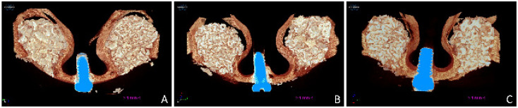
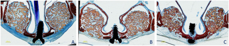
Methods: A maxillary sinus augmentation was performed bilaterally in 18 rabbits using DBBM with particle dimensions of either 0.125-1.0 mm or 1-2 mm. The antrostomy was covered using a collagen barrier. The animals were euthanized in groups of six after 2, 4, and 8 weeks of healing. MicroCT and histological analyses were performed.
Results: After 2 weeks of healing, BGC was 10.9% and 11.9% for the small and large granule sites, respectively. After 8 weeks of healing, the BGC increased to 65% and 62% at the small and large granule sites, respectively. The highest values were located close to the bony walls and the bony window. New bone content developed between 2 and 8 weeks from 7.0% to 27.6% and from 6.1% to 27.6% at the small and large granule sites, respectively.
Conclusions: Similar outcomes in osteoconductivity and bone formation were found at both small and large DBBM granule sites.
Keywords: animal study; bone healing; collagen membrane; histology; morphometry; osteoconductivity; sinus floor elevation; xenograft.
Conflict of interest statement
The authors declare no conflict of interest.
Figures


Similar articles
-
Comparison of histomorphometry and microCT after sinus augmentation using xenografts of different particle sizes in rabbits.Oral Maxillofac Surg. 2020 Mar;24(1):57-64. doi: 10.1007/s10006-019-00813-x. Epub 2019 Dec 9. Oral Maxillofac Surg. 2020. PMID: 31820198
-
Reposition of the bone plate over the antrostomy in maxillary sinus augmentation: A histomorphometric study in rabbits.Clin Oral Implants Res. 2018 Aug;29(8):821-834. doi: 10.1111/clr.13292. Epub 2018 Jun 7. Clin Oral Implants Res. 2018. PMID: 29876969
-
Sequential healing of the elevated sinus floor with different size of antrostomy: a histomorphometric study in rabbits.Oral Maxillofac Surg. 2020 Dec;24(4):403-410. doi: 10.1007/s10006-020-00859-2. Epub 2020 Jun 13. Oral Maxillofac Surg. 2020. PMID: 32535762
-
The Influence on Healing of Bony Window Elevated Inward in the Sinus Cavity as Cortical Bone Graft: A Histomorphometric Study in Rabbit Model.Int J Oral Maxillofac Implants. 2020 Sep/Oct;35(5):879-887. doi: 10.11607/jomi.8226. Int J Oral Maxillofac Implants. 2020. PMID: 32991637
-
Healing at implants installed concurrently to maxillary sinus floor elevation with Bio-Oss® or autologous bone grafts. A histo-morphometric study in rabbits.Clin Oral Implants Res. 2017 May;28(5):503-511. doi: 10.1111/clr.12825. Epub 2016 Mar 10. Clin Oral Implants Res. 2017. PMID: 26969193
Cited by
-
Application of the break-even point to express the bone dynamics around implants.Oral Maxillofac Surg. 2024 Jun;28(2):605-612. doi: 10.1007/s10006-023-01176-0. Epub 2023 Aug 17. Oral Maxillofac Surg. 2024. PMID: 37589916
-
Critical-sized marginal defects around implants treated with xenografts in rabbits.Oral Maxillofac Surg. 2024 Jun;28(2):827-838. doi: 10.1007/s10006-024-01216-3. Epub 2024 Jan 29. Oral Maxillofac Surg. 2024. PMID: 38285089
-
Analysis of Implant Osseointegration, Bone Repair, and Sinus Mucosa Integrity Using Bio-Oss® and Hyaluronic Acid-Polynucleotide Gel (Regenfast®) in Maxillary Sinus Augmentation in Rabbits.Dent J (Basel). 2025 Jun 28;13(7):293. doi: 10.3390/dj13070293. Dent J (Basel). 2025. PMID: 40710138 Free PMC article.
-
Influence on Implant Bone Healing of a Collagen Membrane Placed Subjacent the Sinus Mucosa-A Randomized Clinical Trial on Sinus Floor Elevation.Dent J (Basel). 2022 Jun 8;10(6):105. doi: 10.3390/dj10060105. Dent J (Basel). 2022. PMID: 35735646 Free PMC article.
-
Clinical and Histological Healing after Maxillary Sinus Floor Elevation.Dent J (Basel). 2022 Jul 15;10(7):134. doi: 10.3390/dj10070134. Dent J (Basel). 2022. PMID: 35877408 Free PMC article.
References
-
- Scala A., Botticelli D., Faeda R.S., Garcia Rangel I., Jr., de Oliveira J.A., Lang N.P. Lack of influence of the Schneiderian membrane in forming new bone apical to implants simultaneously installed with sinus floor elevation: An experimental study in monkeys. Clin. Oral Implant. Res. 2012;23:175–181. doi: 10.1111/j.1600-0501.2011.02227.x. - DOI - PubMed
LinkOut - more resources
Full Text Sources
