Review
. 2021 May 31;8(6):461.
doi: 10.3390/children8060461.

Deformity Reconstruction Surgery for Tibial Hemimelia

Affiliations
Review

Deformity Reconstruction Surgery for Tibial Hemimelia

David Y Chong et al. Children (Basel). .

Abstract

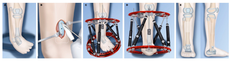
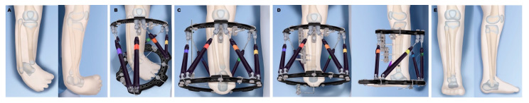
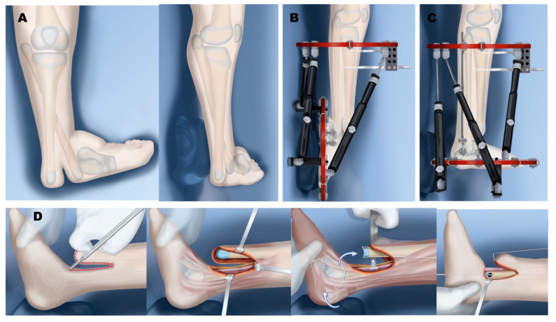
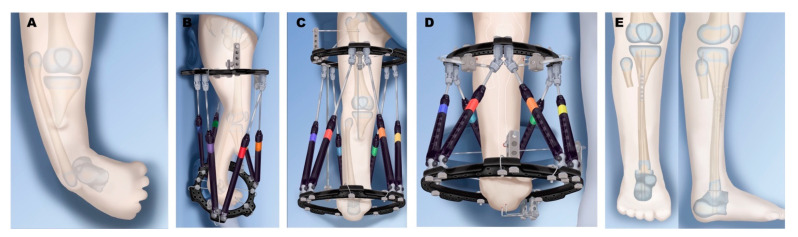
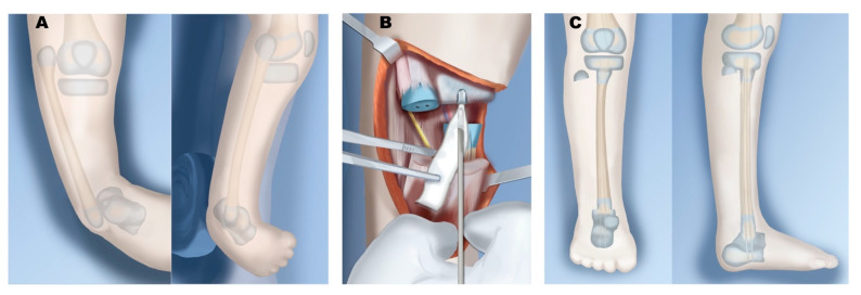
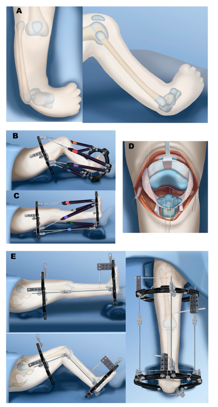
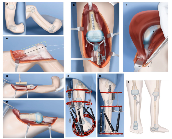
Tibial hemimelia is a rare congenital deficiency with a wide spectrum of pathology and deformity. This paper aims to give a comprehensive review of tibial hemimelia, with a concise summary of the history, pathology, and clinical findings of tibial hemimelia, while providing treatment recommendations and a review of the current literature. Classifications and surgical treatments are discussed, including amputation, limb reconstruction, and lengthening. Type-specific treatments are also discussed, including staged distraction correction of joint contractures of knee and ankle, Weber patelloplasty, fibular centralization, knee and ankle arthrodesis, implantable articulated distractors, and the role of femoral shortening. Amputation is a simpler and easier solution for many patients; however, reconstruction options continue to evolve, improve, and provide better functional outcomes in many cases. Factors favoring surgical reconstruction include the presence of a knee joint/proximal tibia, and the presence of a patella and quadriceps mechanism.

Keywords: Paley classification; absence of tibia; fibula centralization; patelloplasty; tibial aplasia; tibial deficiency; tibial hemimelia; treatment.


Conflict of interest statement

The authors declare no relevant conflict of interest.

Figures

Figure 1
Figure 1
Jones classification of tibial hemimelia.
Figure 2
Figure 2
Paley classification of tibial hemimelia.
Figure 3
Figure 3
Treatment of Paley type 2A. (A) Typical deformity with shortened tibia and equinovarus foot and overgrown proximal fibula. (B) Application of external fixator for staged correction of foot equinovarus, distal fibular transport, and finally lengthening of the tibia. (C) Results after tibial lengthening with distal fibular screw epiphysiodesis.
Figure 4
Figure 4
Treatment of Paley type 2B. (A) Bracket epiphysis deformity of tibia. (B) Application of external fixator after excision of bracket, with staged gradual foot correction, distal fibular transport, and tibial osteotomy for lengthening. (C) Results after completion. The foot has been corrected to a plantigrade position, the fibula is at station with a distal epiphysiodesis, and the tibia is longer due to lengthening.
Figure 5
Figure 5
Treatment of Paley type 2C. (A) Distal tibia cartilaginous anlage with no physis. (B) Insertion of bone morphogenic protein (BMP) into tibial anlage. (C) External fixator for correction of foot deformity and distal fibular transport. (D) Tibial osteotomy and lengthening with fixator. (E) Final results after tibial lengthening with fibula at station and foot plantigrade.
Figure 6
Figure 6
Treatment of Paley type 3A. (A) Distal tibia-fibula diastasis. The tibial plafond is absent and the end of the tibia is what normally would have been a medial malleolus; foot and fibula internally rotated around tibia. (B) Application of external fixator for gradual correction of foot and fibula position. (C) Foot centralized under end of tibia, ready for tibiotalar arthroplasty. (D) Distal tibia reshaping to talus and stabilization of distal tibia and fibula diastasis and osteotomy of tibia for diaphyseal straightening. (E) Final result after hardware removal with plantigrade foot and distal fibula screw epiphysiodesis.
Figure 7
Figure 7
Treatment of Paley type 3B. (A) Distal tibia and fibula diastasis with skin cleft. Fibula is associated with talus and foot. (B) Application of external fixator for distraction and gradual correction. (C) Foot now in plantigrade position. (D) Excision and closure of skin cleft performed at time of diastasis stabilization and tibiotalar biologic arthroplasty as in type 3A.
Figure 8
Figure 8
Treatment of Paley type 4A. (A) Well-formed proximal tibia and knee with distal tibia aplasia. (B) External fixator used to gradually correct equinovarus foot position and distally transport fibular head. (C) Fibula brought down to station and foot plantigrade. (D) Fibular osteotomy performed with transfer to proximal tibia. Distal fibula epiphysis is fused to talus without disrupting the physis. Fusion stabilized with intramedullary retrograde wires. (E) Final results after fixator removal.
Figure 9
Figure 9
Treatment of Paley type 4B. (A) Unossified proximal tibial anlage with no physis. Initial treatment the same as 4A, with fibula distraction and foot correction. (B) Insertion of BMP into proximal tibia anlage. (C) Fixator removal following healing from physeal sparing proximal fibula osteotomy and transfer to proximal tibia. Physeal-sparing fusion of distal fibula to talus.
Figure 10
Figure 10
Treatment of Paley type 5A. (A) Complete aplasia of tibia, but patella and quadriceps are present. (B) External fixator placement for gradual fibula distraction and foot correction. (C) Knee flexion contracture corrected to straight position, preparing for physeal sparing patellar arthroplasty and physeal sparing talo-fibular fusion. (D) Paley-Weber patelloplasty converting the patella into a tibial plateau. (E) Hinged external fixator to protect arthroplasty but allow knee motion.
Figure 11
Figure 11
Treatment of Paley type 5C. (A) Complete aplasia of tibia with no patella. (B) Quadriceps are distally absent and end in distal femur. Femur needs to be shortened significantly to be able to bring the quadriceps to the level of the knee joint. (C) Femoral shortening osteotomy. (D) Plate fixation and intramedullary pinning of femoral osteotomy and distalization and centralization of fibula. (E) Knee stabilized with IJS internal articulated joint distractor. Collateral ligaments made from autograft or allograft. Reconstruction of collateral ligaments and placement of internal joint distraction system. (F) After quadricepsplasty, quadriceps muscle is advanced and sutured to the fibula head. (G) Placement of two-level external fixator: upper two rings with hinges for articulated stabilization of knee; lower two rings for gradual foot distraction. (H) Once talus is beneath the fibula and foot at 90°, physeal sparing fusion of foot to talus is carried out. (I) Final results after fixator removal. The IJS device side arm is disconnected 6 months later to allow for proximal fibular growth.

Similar articles

Cited by

References

    1. Brown F.W. The Brown Operation for Total Hemimelia Tibia. In: Aitken G.T., editor. Selected Lower-Limb Anomalies: Surgical and Prosthetics Management: A Symposium Held in Washington. The National Academies Press; Washington, DC, USA: 1971.
    1. Weber M., Schroder S., Berdel P., Niethard F.U. Nation-wide registration of limb deficiencies in Germany. Z. Orthop. Ihre. Grenzgeb. 2005;143:534–538. doi: 10.1055/s-2005-872467. - DOI - PubMed
    1. Otto A.W. Monstrorum sexcentorum descriptio anatomica. Vratislaviae (Breslau): Sumptibus Ferdinandi Hirt, 1841. Print.
    1. Billroth T. Ueber einige durch Knochendefecte bedingte Krümmungen des Fusses. Arch Klin. Chir. 1861;1:252–268.
    1. Dankmeijer J. Congenital absence of the tibia. Anat. Rec. 1935;62:179194. doi: 10.1002/ar.1090620205. - DOI

LinkOut - more resources