. 2021 May 31;22(11):5916.
doi: 10.3390/ijms22115916.

Bio-Functionalized Chitosan for Bone Tissue Engineering

Affiliations

Bio-Functionalized Chitosan for Bone Tissue Engineering

Paola Brun et al. Int J Mol Sci. .

Abstract

Hybrid biomaterials allow for the improvement of the biological properties of materials and have been successfully used for implantology in medical applications. The covalent and selective functionalization of materials with bioactive peptides provides favorable results in tissue engineering by supporting cell attachment to the biomaterial through biochemical cues and interaction with membrane receptors. Since the functionalization with bioactive peptides may alter the chemical and physical properties of the biomaterials, in this study we characterized the biological responses of differently functionalized chitosan analogs. Chitosan analogs were produced through the reaction of GRGDSPK (RGD) or FRHRNRKGY (HVP) sequences, both carrying an aldehyde-terminal group, to chitosan. The bio-functionalized polysaccharides, pure or "diluted" with chitosan, were chemically characterized in depth and evaluated for their antimicrobial activities and biocompatibility toward human primary osteoblast cells. The results obtained indicate that the bio-functionalization of chitosan increases human-osteoblast adhesion (p < 0.005) and proliferation (p < 0.005) as compared with chitosan. Overall, the 1:1 mixture of HVP functionalized-chitosan:chitosan is the best compromise between preserving the antibacterial properties of the material and supporting osteoblast differentiation and calcium deposition (p < 0.005 vs. RGD). In conclusion, our results reported that a selected concentration of HVP supported the biomimetic potential of functionalized chitosan better than RGD and preserved the antibacterial properties of chitosan.

Keywords: Chit-HVP; Chit-RGD; NEXAFS; XPS; chitosan; functionalization; h-osteoblasts.


Conflict of interest statement

The authors declare no conflict of interest.

Figures

Figure 1
Figure 1
C1s (A), O1s (B) and N1s (C) spectra of RGD peptide and functionalized chitosan matrix Chit-RGD deposited on titania and curve-fitting analysis of the experimental spectra.
Figure 2
Figure 2
C1s (A), O1s (B) and N1s (C) spectra of HVP peptide and Chit-HVP functionalized chitosan matrix deposited on titania and curve-fitting analysis of the experimental spectra.
Figure 3
Figure 3
Carbon K-edge spectra of RGD and Chit-RGD (A) and of HVP and Chit-HVP (B).
Figure 4
Figure 4
Nitrogen K-edge spectra of RGD and Chit-RGD (A) and of HVP and Chit-HVP (B).
Figure 5
Figure 5
Angular dependent N K-edge NEXAFS spectra collected at normal (90°) and grazing (20°) incidence angles of the impinging X-ray beam on (A) RGD; (B) HVP; (C) Chit-RGD; (D) Chit-HVP. The difference spectra (grazing-normal), evidencing dichroic effects, are also shown (green lines).
Figure 6
Figure 6
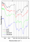
FTIR spectra of the pristine peptides (HVP, RGD) and the corresponding chitosan-peptide assemblies (Chit-HVP, Chit-RGD) in the 3900–2600 and 1900–600 cm−1 wavenumber ranges.
Figure 7
Figure 7
MTT assay performed in primary human osteoblast cells seeded for 2 h on different functionalized chitosan matrices. The cells were quantified by setting a standard curve for each experiment obtained by planting a known number of osteoblast cells. In (A), the percentage of cell survival was calculated on the number of cells retrieved in chitosan. (B,C) number of cells adhering to different functionalized chitosan. Data are reported as mean ± st err of three independent experiments, each performed in duplicate. * indicates p < 0.005. ** indicates p < 0.001. PCS: plastic culture surface; Chit: chitosan. % indicates the concentration of HVP or RGD.
Figure 8
Figure 8
Immunofluorescence on human primary osteoblast cells cultured for 24 h on differently functionalized chitosan matrices. Cells were stained with anti-αSMA antibody (red). Nuclei were stained with TOTO-3 (blue). The images were acquired using a Leica TCSNT/SP2 confocal microscope (Leica Microsystems). Scale bar: 37.5 μm. The images are representative of three independent experiments.
Figure 9
Figure 9
Cell proliferation evaluated in primary human osteoblast cells loaded with CFSE intracellular fluorescent dye. The cells were cultured for 72 h on chitosan matrices and then analyzed by cytofluorimetric analysis. Data are reported as mean ± st err of three independent experiments, each performed in duplicate. * indicates p < 0.005. PCS: plastic culture surface; Chit: chitosan. % indicates the concentration of HVP or RGD.
Figure 10
Figure 10
Quantitative RT-PCR performed on human osteoblast cells cultured for 24 h on functionalized chitosan matrices. Data were quantified by the ΔΔCT method using hGAPDH as a reference gene. Data are reported as mean ± st err of three independent experiments, each performed in triplicate. * indicates p < 0.005 vs. Chit. PCS: plastic culture surface; Chit: chitosan. % indicates the concentration of HVP or RGD.
Figure 11
Figure 11
Osteoblast cells were cultured for 7 days on functionalized chitosan matrices and then fixed and stained with Alizarin red to visualize calcium deposition. (A) The images were acquired using a Leica microscope equipped with a digital camera. (B) The cells were lysed in acetic acid, and the optical density (O.D.) was recorded at 405 nm. Data are reported as the mean ± st err of three independent experiments, each performed in duplicate. * indicates p < 0.005 vs. Scale bar: 75 μm. Chit. PCS: plastic culture surface; Chit: chitosan. % indicates the concentration of HVP or RGD.
Figure 12
Figure 12
Cultures of S. aureus were grown on different functionalized chitosan matrices for 48 h, resulting in a mature biofilm in glass surfaces. Bacterial cultures were stained with a LIVE/DEAD BacLight Bacterial Viability Kit and (A) images were acquired using a Leica TCSNT/SP2 confocal microscope. Scale bar: 37.5 μm. The images are representative of three independent experiments, each performed in duplicate. (B) Data were analyzed using ImageJ and the ratio of live (bacteria with SYTO9 green fluorescence)/dead (bacteria with a high propidium iodide fluorescence and a low SYTO9 green fluorescence) bacteria was recorded. Data are reported as mean ± st err. * indicates p < 0.005 vs. glass surfaces. Chit: chitosan. % indicates the concentration of HVP or RGD.

References

    1. Bagheri-Khoulenjani S., Taghizadeh S., Mirzadeh H. An Investigation on the Short-Term Biodegradability of Chitosan with Various Molecular Weights and Degrees of Deacetylation. Carbohydr. Polym. 2009;78:773–778. doi: 10.1016/j.carbpol.2009.06.020. - DOI
    1. Zhang Y., Sun T., Jiang C. Biomacromolecules As Carriers in Drug Delivery and Tissue Engineering. Acta Pharm. Sin. B. 2018;8:34–50. doi: 10.1016/j.apsb.2017.11.005. - DOI - PMC - PubMed
    1. Palma P.J., Ramos J., Martins J.B., Diogenes A., Figueiredo M.H., Ferreira P., Viegas C., Santos J.M. Histologic Evaluation of Regenerative Endodontic Procedures with the Use of Chitosan Scaffolds in Immature Dog Teeth with Apical Periodontitis. J. Endod. 2017;43:1279–1287. doi: 10.1016/j.joen.2017.03.005. - DOI - PubMed
    1. Huang Y.-M., Lin Y.-C., Chen C.-Y., Hsieh Y.-Y., Liaw C.-K., Huang S.-W., Tsuang Y.-H., Chen C.-H., Lin F.-H. Thermosensitive Chitosan–Gelatin–Glycerol Phosphate Hydrogels as Collagenase Carrier for Tendon–Bone Healing in a Rabbit Model. Polymers. 2020;12:436. doi: 10.3390/polym12020436. - DOI - PMC - PubMed
    1. Hansson A., Hashom N., Falson F., Rousselle P., Olivier J., Borchard E.G. In Vitro Evaluation of an RGD-Functionalized Chi-Tosan Derivative for Enhanced Cell adhesion. Carbohydr. Polym. 2012;90:1494–1500. doi: 10.1016/j.carbpol.2012.07.020. - DOI - PubMed

MeSH terms