Review
. 2021 May 31;11(6):823.
doi: 10.3390/biom11060823.

Understanding Emotions: Origins and Roles of the Amygdala

Affiliations
Review

Understanding Emotions: Origins and Roles of the Amygdala

Goran Šimić et al. Biomolecules. .

Abstract

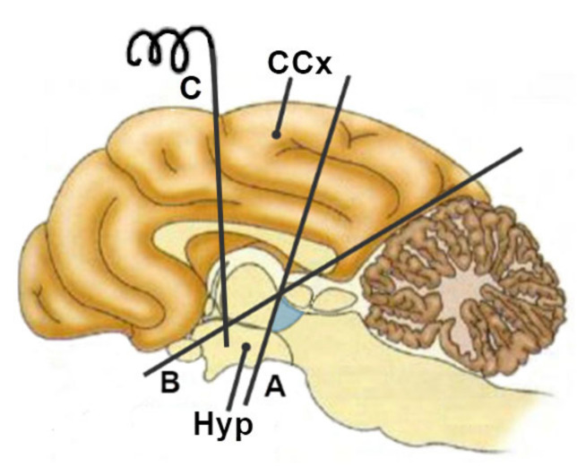
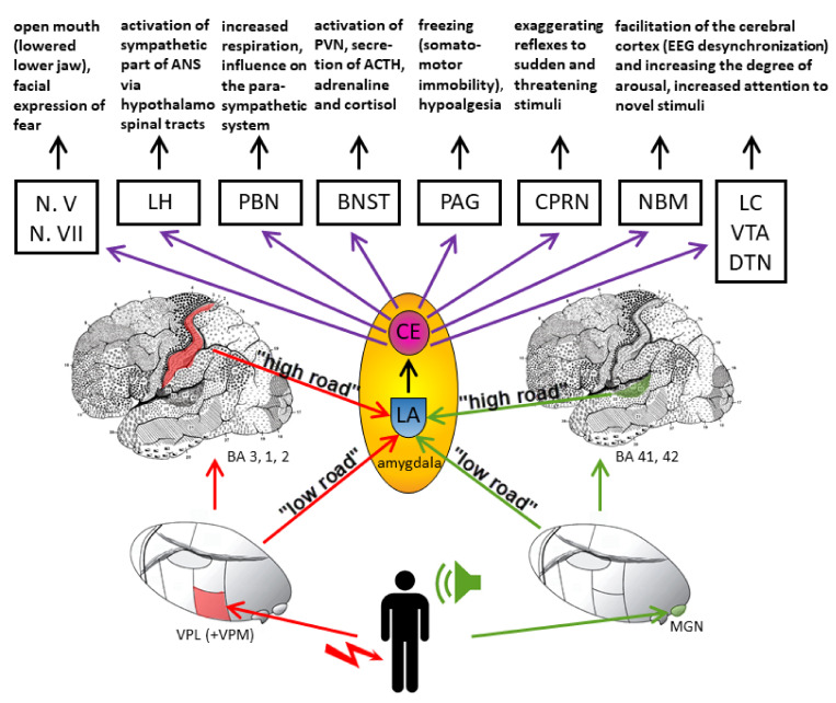
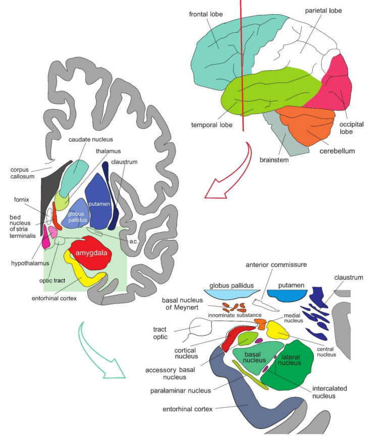
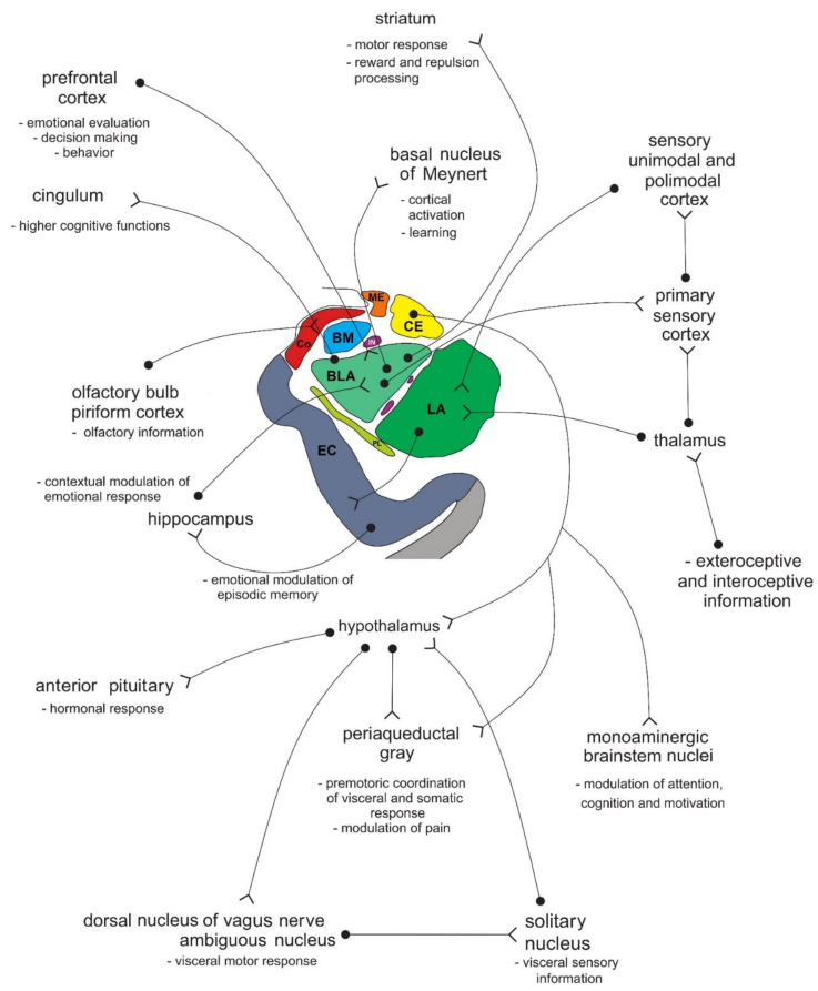
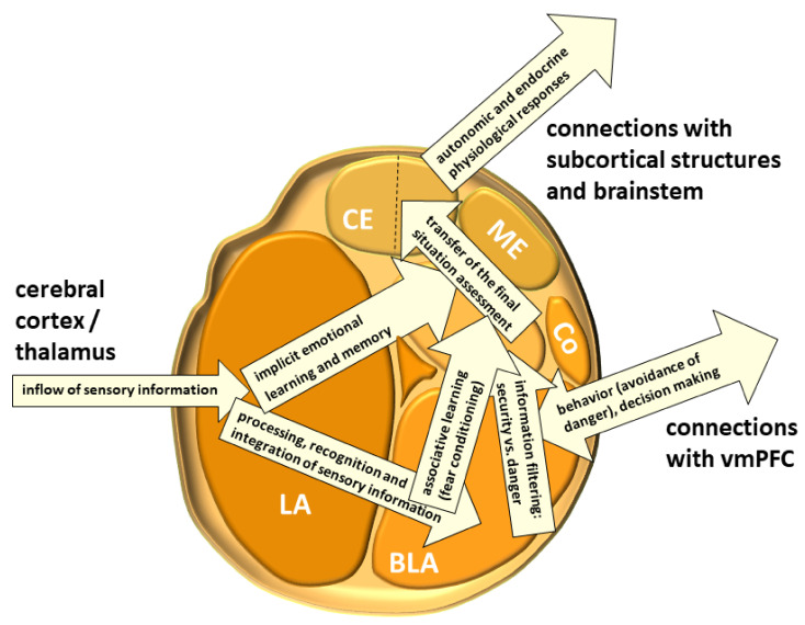
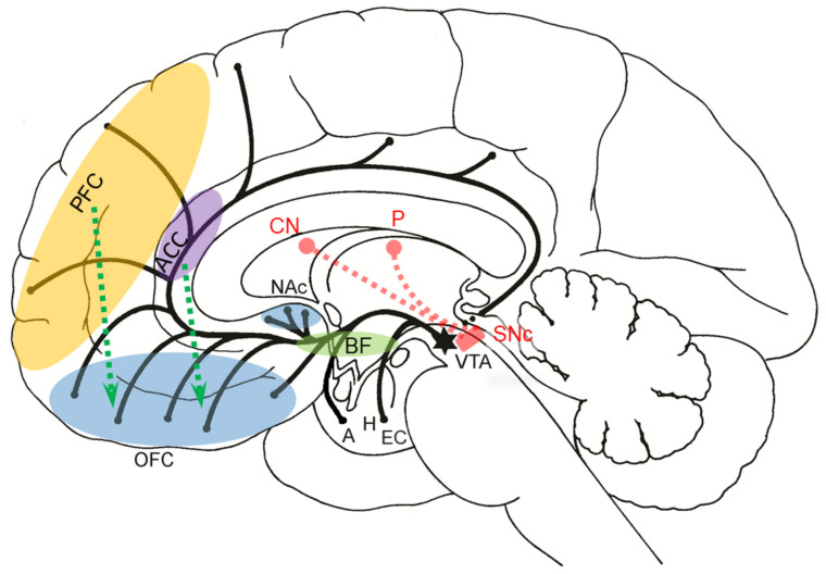
Emotions arise from activations of specialized neuronal populations in several parts of the cerebral cortex, notably the anterior cingulate, insula, ventromedial prefrontal, and subcortical structures, such as the amygdala, ventral striatum, putamen, caudate nucleus, and ventral tegmental area. Feelings are conscious, emotional experiences of these activations that contribute to neuronal networks mediating thoughts, language, and behavior, thus enhancing the ability to predict, learn, and reappraise stimuli and situations in the environment based on previous experiences. Contemporary theories of emotion converge around the key role of the amygdala as the central subcortical emotional brain structure that constantly evaluates and integrates a variety of sensory information from the surroundings and assigns them appropriate values of emotional dimensions, such as valence, intensity, and approachability. The amygdala participates in the regulation of autonomic and endocrine functions, decision-making and adaptations of instinctive and motivational behaviors to changes in the environment through implicit associative learning, changes in short- and long-term synaptic plasticity, and activation of the fight-or-flight response via efferent projections from its central nucleus to cortical and subcortical structures.

Keywords: amygdala; anxiety; emotion; evolution; fear.


Conflict of interest statement

The authors declare no conflict of interest.

Figures

Figure 1
Figure 1
Emotional facial expressions of three basic, primary emotions. At the top is a neutral facial expression. In the bottom row, facial expressions of anger, joy, and fear are shown, respectively. Although individual emotions can be recognized and analyzed even from the microexpressions of facial muscles, for the sake of clarity the expressions of emotions in these photographs are accentuated. See text for details. Photographs by Andrea Piacquadio, taken from [9].
Figure 2
Figure 2
Schematic representation of Bard’s experiments on cats. Behavior described as “false anger/false rage” occurs if the cutting line when decorticating a cat goes from the posterior part of the cerebral cortex through the anterior part of the hypothalamus (line marked with (B)), but not if it goes through its posterior part (line marked with (A)). In both cases, a small part of the caudoventral part of the thalamus remains preserved (marked in blue). Electrical stimulation of the hypothalamus with an electrode (without cutting) leads to anger and fear (C). The schematic follows Bard’s textual description (Bard, 1928) [35]. CCx—cerebral cortex; Hyp—hypothalamus. See text for details.
Figure 3
Figure 3
Simplified schematic representation of classical theories of emotion. Photographs taken from [9,48]. ANS—autonomic nervous system. See text for details.
Figure 4
Figure 4
Simplified schematic representation of neural circuits underlying fear conditioning. Pathways that process a conditioned stimulus (CS, auditory pathway, green) and an unconditioned stimulus (US, spinothalamic anterolateral pain pathway, red) via the ventroposterolateral (VPL) and ventroposteromedial (VPM) nuclei and the medial geniculate body (MGN) of the thalamus monosynaptically, and via the cerebral cortex of Brodmann’s areas 3, 1, and 2 (primary somatosensory cortex); 41 and 42 (primary auditory cortex) polysynaptically converge on the lateral nucleus of the amygdala (LA, the LA receives the majority of afferent fibers). CS-US convergence in LA initiates long-term potentiation (LTP), leading to the creation of a learned association between the two stimuli. LA activity is then transferred to the central nucleus (CE, the central nucleus of the amygdala), which sends most of the efferent projections to a number of different cortical and subcortical areas through which the amygdala directly regulates autonomic responses and context-dependent behavior: ANS, reflexes, and hormone secretion. Sympathetic activation includes mydriasis, tachycardia, hypertension, peripheral vasoconstriction, cessation of peristalsis, sphincter contraction, and other effects. All these effects help organisms to cope with threat. Synaptic plasticity also changes in neurons in other nuclei of the amygdala (intentionally omitted here). ACTH—adrenocorticotropic hormone; BA—Brodmann’s area; BNST—bed nucleus of stria terminalis; CPRN—caudal pontine reticular nucleus; DTN—dorsal tegmental nucleus; EEG—electroencephalogram; LC—locus coeruleus; LH—lateral hypothalamus; MGN—medial geniculate nucleus; NBM—nucleus basalis Meynerti; N. V—trigeminal nerve; N. VII—facial nerve; PAG—periaqueductal gray; PBN—parabrachial nuclei; PVN—paraventricular nucleus; VPL and VPM—ventroposterolateral and ventroposteromedial thalamic nuclei; VTA—ventral tegmental area. The schematic is made according to LeDoux [73,74].
Figure 5
Figure 5
Simplified representation of the structure and location of the amygdala. The upper part of the schematic shows the human brain when viewed from the lateral side, where the brainstem, cerebellum, and four lobes of the cerebrum can be seen. The middle part of the schematic shows the structures present on the coronal plane through the temporal lobe of the cerebrum on which the position of the amygdala can be observed. The lower part of the schematic shows an enlarged amygdala with its individual nuclei. a.c.—anterior commissure. See text for details.
Figure 6
Figure 6
Simplified schematic representation of the connections of individual amygdala nuclei with numerous cortical and subcortical structures, and their role in processing functionally different types of information. Amygdala nuclei are marked in colors as shown in Figure 5. BLA—basolateral (basal) nucleus; BM—basomedial (accessory basal) nucleus; CE—central nucleus; Co—cortical nucleus; EC—entorhinal cortex; IN—intercalated neurons; ME—medial nucleus; LA—lateral nucleus; PL—paralaminar nucleus. See text for details.
Figure 7
Figure 7
Simplified neuroanatomical representation of information flow within the amygdala. BLA—basolateral nucleus of the amygdala; CE—central nucleus of the amygdala; Co—cortical nucleus of amygdala; IN—intercalate neurons; LA—lateral nucleus of amygdala; ME—medial nucleus of amygdala; BM—basomedial (accessory basal) nucleus of the amygdala. The schematics is made according to Wieronska et al., (2010) [148], Orsini and Maren (2012) [111], Benarroch (2015) [149], Gilpin et al., (2015) [116], Janak and Tye (2015) [147], and Sangha et al., (2020) [80].
Figure 8
Figure 8
Balanced ratio of excitation and inhibition in amygdala in a healthy individual in a non-threatening situation. AMPA—α-amino-3-hydroxy-5-methyl-4-isoxazole propionic acid; BLA—basolateral nucleus of the amygdala; CE—central nucleus of the amygdala; GABA—γ-aminobutyric acid; IN—intercalated neurons; LA—lateral nucleus of amygdala; NMDA—N-methyl-D-aspartate.
Figure 9
Figure 9
Schematic representation of the predominance of excitation over inhibition in circumstances of imminent danger, but also in anxiety and other functional disorders of the amygdala. The central nucleus of the amygdala contains different populations of GABAergic neurons. This area mediates inhibitory control over the lateral region of the amygdala [111]. AMPA—α-amino-3-hydroxy-5-methyl-4-isoxazole propionic acid; BLA—basolateral nucleus of the amygdala; CE—central nucleus of the amygdala; GABA—γ-aminobutyric acid; IN—intercalated neurons; LA—lateral nucleus of amygdala; NMDA—N-methyl-D-aspartate.
Figure 10
Figure 10
Simplified representation of the information flow within the amygdala. BLA—basolateral nucleus of the amygdala; CE—central nucleus of the amygdala; Co—cortical nucleus of amygdala; LA—lateral nucleus of amygdala; ME—medial nucleus of amygdala; vmPFC—the ventromedial prefrontal cortex; BM—basomedial (accessory basal) nucleus of the amygdala. The schematics are made according to Sah et al., (2017) [108], Asami et al., (2018) [162], and Neugebauer (2020) [135].
Figure 11
Figure 11
Ontogenesis of individual primary and secondary emotions in the first two years of life. According to Banham Bridges (1932) [207].
Figure 12
Figure 12
Schematic representation of the development of select emotional and cognitive abilities in children. ACC—anterior cingulate cortex; vmPFC—ventromedial prefrontal cortex. The part of the schematic related to the stages of development is made according to Lewis and Granic (2010) [209].
Figure 13
Figure 13
Schematic representation of dopaminergic projections that make up the brain reward system. The projections originate from the neurons of the ventral tegmental area (VTA, black star) and go to the ventral striatum (ventral pallidum), especially the nucleus accumbens septi (NAc, small blue ellipse, mesolimbic pathway), orbitofrontal cortex (OFC, large blue ellipse) and prefrontal cortex (PFC, yellow ellipse, mesocortical pathway), anterior cingulate cortex (ACC, purple ellipse) and mediobasal telencephalon (basal forebrain, BF, green ellipse), entorhinal cortex (EC), hippocampus (H) and amygdala (A). The release of dopamine from projecting VTA neurons in other parts of the CNS, especially the hippocampus (H) and the amygdaloid nucleus (A) is associated with the memory of (otherwise neutral) individual stimuli/objects/events present during rewarding, which gives them motivational importance [256,257,258]. It is thought that dopaminergic projections from the substantia nigra, pars compacta (SNc, red rectangle) to the dorsal striatum, i.e., caudate nucleus (CN) and putamen (P) also transmit information that associate salient sensory stimuli with reward and reward prediction error, but in this context they are associated with the dopaminergic “tone” necessary to perform conscious motor movements and to reprogram motor patterns that will facilitate obtaining the same reward in the future [259]. Green dashed arrows represent projections of the PFC and ACC in the OFC. These projections are thought to exert cognitive (top-down) control over glutamatergic and GABAergic interactions in the OFC, a key region responsible for making behavioral choices, such as emotional go/no-go decisions. Schematic modified from Šešo-Šimić et al., 2010 [169].
Figure 14
Figure 14
Schematic drawing of the main sites and mechanisms of action of some common addictive drugs on the brain reward system: reward learning and motivation are strongly influenced by the amygdala. Thick blue arrows from OFC, AMY, and HF to NAc convey contextual information associated with the addictive substance and contribute to relapse. Although many addictive substances directly stimulate the release of dopamine from neurons in the VTA that are projected into the NAc, it must not be forgotten that the same effect (activation of VTA) with drug-related stimuli can be achieved indirectly through projections from the amygdala to the PFC and then from PFC to VTA [265]. In a state of developed dependence, the reward system is active, but the usual (normal) reward can no longer activate it. This state of motivational toxicity is expressed in hardened addicts. It is manifested by a lack of interest in career, social and sexual relations, financial status and increased engagement in the procurement and consumption of drugs. The diagram does not show the efferent projections of NAc that go to the basal ganglia and ventral pallidum. Neurons of the ventral pallidum are projecting through the mediodorsal nucleus of the thalamus into the PFC and striatum, and additional projections go into the RMTg, the compact part of the substantia nigra (SNc), and the reticular formation of the pons. Not shown are glutamatergic projections from the thalamus and ACC into Nac, as well as projections of NAc and ventral pallidum into the lateral hypothalamus. AMPA—α-amino-3-hydroxy-5-methyl-4-isoxazolepropionic acid; AMY—amygdala; AP-1—transcription factor activating protein 1; ATP/Ado—adenosine triphosphate/adenosine; BLA—basolateral nucleus of amigdala; DA—dopamine; DRN—dorsal raphe nucleus; ΔFosB & JUN—truncated member of the Fos family of transcription factors and JUN protein (ΔFosB↑* in RMTg applies only to psychostimulants); GABA—γ-aminobutyric acid; GLU—glutamate; HF—hippocampal formation; LTD—long-term depression; MDMA—3,4-methylenedioxymethamphetamine (ecstasy); NAc MSN—medium spiny neurons in nucleus accumbens septi; OFC—orbitofrontal cortex; RMTg—rostromedial tegmental nucleus; VSu—ventral subiculum; VTA—ventral tegmental area. See text for details.

References

    1. Vingerhoets A., Nykliček I., Denollett J. Emotion Regulation: Conceptual and Clinical Issues. Springer; New York, NY, USA: 2008.
    1. Gračanin A., Kardum I. Primary emotions as modular mechanisms of the human mind. In: Žebec M.S., Sabol G., Šakić M., Topić M.K., editors. Brain and Mind: A Lasting Challenge. Institute of Social Sciences “Ivo Pilar”; Zagreb, Croatia: 2006. pp. 89–103.
    1. Fox E. Emotion Science. J.B. Metzler; Stuttgart, Germany: 2008.
    1. Adolphs R., Anderson D.J. The Neuroscience of Emotion: A New Synthesis. Princeton University Press; Princeton, NJ, USA: 2018.
    1. Ekman P. An argument for basic emotions. Cogn. Emot. 1992;6:169–200. doi: 10.1080/02699939208411068. - DOI

Publication types

LinkOut - more resources