. 2021 May 31;9(6):1194.
doi: 10.3390/microorganisms9061194.

Innovative Approach to Fast Electron Microscopy Using the Example of a Culture of Virus-Infected Cells: An Application to SARS-CoV-2

Affiliations

Innovative Approach to Fast Electron Microscopy Using the Example of a Culture of Virus-Infected Cells: An Application to SARS-CoV-2

Marion Le Bideau et al. Microorganisms. .

Abstract

Despite the development of new diagnostic methods, co-culture, based on sample inoculation of cell monolayers coupled with electron microscopy (EM) observation, remains the gold standard in virology. Indeed, co-culture allows for the study of cell morphology (infected and not infected), the ultrastructure of the inoculated virus, and the different steps of the virus infectious cycle. Most EM methods for studying virus cycles are applied after infected cells are produced in large quantities and detached to obtain a pellet. Here, cell culture was performed in sterilized, collagen-coated single-break strip wells. After one day in culture, cells were infected with SARS-CoV-2. Wells of interest were fixed at different time points, from 2 to 36 h post-infection. Microwave-assisted resin embedding was accomplished directly in the wells in 4 h. Finally, ultra-thin sections were cut directly through the infected-cell monolayers. Our methodology requires, in total, less than four days for preparing and observing cells. Furthermore, by observing undetached infected cell monolayers, we were able to observe new ultrastructural findings, such as cell-cell interactions and baso-apical cellular organization related to the virus infectious cycle. Our innovative methodology thus not only saves time for preparation but also adds precision and new knowledge about viral infection, as shown here for SARS-CoV-2.

Keywords: SARS-CoV-2; electron microscopy; embedding method; microwaves; single-break strip.


Conflict of interest statement

The authors declare no conflict of interest.

Figures

Figure 1
Figure 1
Schematic representation of the methodology for fast electron microscopic examination of a virus culture. (1) Removable single-break strips are UV sterilized and collagen-coated. (2) Cell culture with 200 µL suspensions of Vero E6 cells at 2 × 10⁵ cells/mL per well. (3) Cells are virus-inoculated with 50 µL of strains or clinical sample per well. (4) Cytopathic effect detection: cytopathic effect (4a) is monitored by bright-field light microscopy, in comparison with a negative control (4b). (5) Cell fixation: cell monolayers are fixed with 2.5% final glutaraldehyde. (6) In-well resin embedding in 4 h: washes, dehydration, resin substitution and wells closed by cap sealing for microwave polymerization. (7) Plastic pruning: resin blocks are manually trimmed with a razor blade. (8) Detachment of the cell monolayer: the plastic bottom of the well is detached by cold shock via immersion in liquid nitrogen for 20 s. (9) Resin block: the resin block containing the cell monolayer at one side is ready for ultramicrotomy. (10) Ultramicrotomy: the resin block is trimmed to a pyramid and ultra-thin sections are obtained. (11) Positioning on grids: ultra-thin sections are deposited on copper/rhodium grids. (12) Contrast and metal deposition: sections are contrasted with uranyl acetate and lead citrate and grids attached to a glass slide are platinum sputter-coated. (13) Electron microscopy: electron micrographs are obtained by SEM.
Figure 2
Figure 2
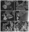
Ultra-thin sections of SARS-CoV-2-infected Vero E6 cells: comparison between images obtained by our classical methodology [9] (left column) and our new approach (right column). In sections obtained from a pellet of SARS-CoV-2-infected cells (A,C), cells were generally isolated and separated from each other, while in sections of SARS-CoV-2 monolayers (B,D), cells were usually clustered, contacting each other, and more spread out. With our new approach, SARS-CoV-2 particles (arrows) could be recognized (F), as was the case when using our classical methodology (E).
Figure 3
Figure 3
Ultra-thin sections of SARS-CoV-2-infected Vero E6 cells: 2–6 hpi. (AC): 2 hpi. Two cells (A) with plasma membranes closely associated with a ‘Velcro’ organization (C; arrowheads). One of the two cells presents a large vacuole enriched in membranes and granules (B). (DI): 3 hpi. One cell (D) presents the typical budding of the endoplasmic reticulum and/or Golgi apparatus compartments following SARS-CoV-2 infection (E,F). (GI): 6 hpi. A region (H) located between intact cells and containing debris and mature SARS-CoV-2 particles (I; arrow).
Figure 4
Figure 4
Ultra-thin sections of SARS-CoV-2-infected Vero E6 cells: 12 hpi. (AC): Two neighboring cells, one with a long filopodia (A,B; arrow A) and one presenting extracellular SARS-CoV-2 particles at the plasma membrane (C; arrow). (DF): SARS-CoV-2-enriched microvilli located between neighboring cells (E; arrows) or at the cell-free periphery (F). (GI): One microvillus extremely enriched in SARS-CoV-2 virions (I; arrow).
Figure 5
Figure 5
Ultra-thin sections of SARS-CoV-2-infected Vero E6 cells: 18 hpi. (A,B): Two serial sections of the same cell, with cuts at the level of its nucleus (*) at different heights along its depth. In (C), the nuclear membrane/endoplasmic reticulum presents a stacked organization (arrow). (DF): Adjacent cells contacting each other through microvilli (E) or electron-dense regions resembling tight junctions (F; arrowheads). (GI): SARS-CoV-2 particles (arrows) located inside a canalicular tubulo-vesicular system (H) as well as in a virus-morphogenesis vesicle (VMV; I) of the same cell.
Figure 6
Figure 6
Ultra-thin sections of SARS-CoV-2-infected Vero E6 cells: 18 hpi. (AD): Neighboring cells harboring extracellular SARS-CoV-2 particles at the level of the intercellular space (C) or at the level of peripheral microvilli (D). (EH): A cellular cluster with two cells facing each other with intermingled microvilli enriched in SARS-CoV-2 particles (H; arrow).
Figure 7
Figure 7
Ultra-thin sections of SARS-CoV-2-infected Vero E6 cells: 36 hpi. (A,B): A cell with a filopodia (B) containing actin microfilaments (arrow); SARS-CoV-2 virions were not detected inside such filopodia structures. (CF): Cells with SARS-CoV-2 particles inside a virus-morphogenesis vesicle (VMV) or inside tubules/vesicles of the canaliculi system (D,E; arrows) or at the plasma membrane (F; arrow). (G,H): Large VMV containing SARS-CoV-2 virions (arrow) and membranes. (IK): In adjacent cellular regions, SARS-CoV-2 particles (arrows) were found concentrated in a VMV, disseminated in a larger vacuole (J; arrows), or stitched extracellularly to the plasma membrane remnants of a lysed cell (K).

Similar articles

Cited by

References

    1. Fong C.K. Electron Microscopy for the Rapid Detection and Identification of Viruses from Clinical Specimens. Yale J. Biol. Med. 1989;62:115–130. - PMC - PubMed
    1. Roingeard P. Viral Detection by Electron Microscopy: Past, Present and Future. Biol. Cell. 2008;100:491–501. doi: 10.1042/BC20070173. - DOI - PMC - PubMed
    1. Goldsmith C.S., Miller S.E. Modern Uses of Electron Microscopy for Detection of Viruses. Clin. Microbiol. Rev. 2009;22:552–563. doi: 10.1128/CMR.00027-09. - DOI - PMC - PubMed
    1. Laue M. Electron Microscopy of Viruses. Methods Cell Biol. 2010;96:1–20. doi: 10.1016/S0091-679X(10)96001-9. - DOI - PubMed
    1. Goldsmith C.S., Ksiazek T.G., Rollin P.E., Comer J.A., Nicholson W.L., Peret T.C.T., Erdman D.D., Bellini W.J., Harcourt B.H., Rota P.A., et al. Cell Culture and Electron Microscopy for Identifying Viruses in Diseases of Unknown Cause. Emerg. Infect. Dis. 2013;19:886–891. doi: 10.3201/eid1906.130173. - DOI - PMC - PubMed

LinkOut - more resources