Review
. 2021 May 31;22(11):5967.
doi: 10.3390/ijms22115967.

Extracellular Vesicles: Versatile Nanomediators, Potential Biomarkers and Therapeutic Agents in Atherosclerosis and COVID-19-Related Thrombosis

Affiliations
Review

Extracellular Vesicles: Versatile Nanomediators, Potential Biomarkers and Therapeutic Agents in Atherosclerosis and COVID-19-Related Thrombosis

Adriana Georgescu et al. Int J Mol Sci. .

Abstract

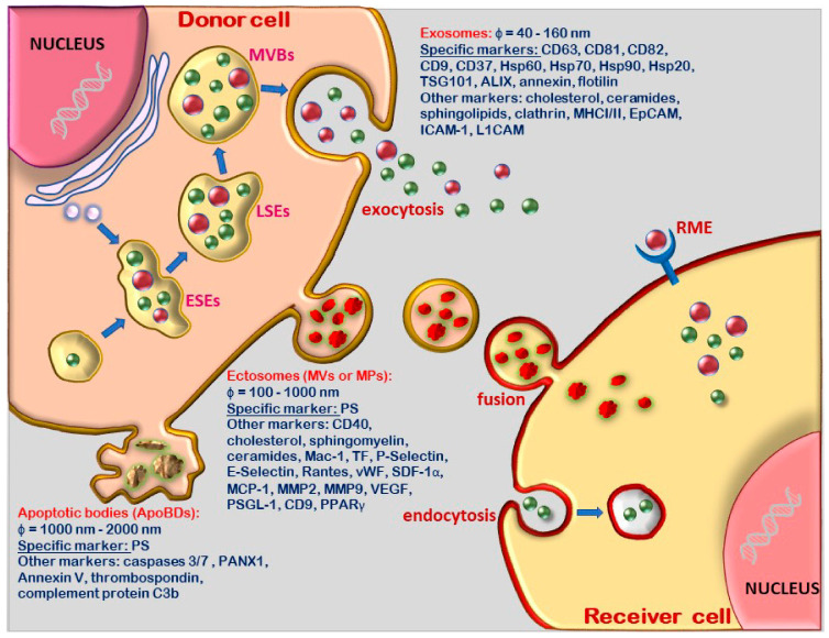
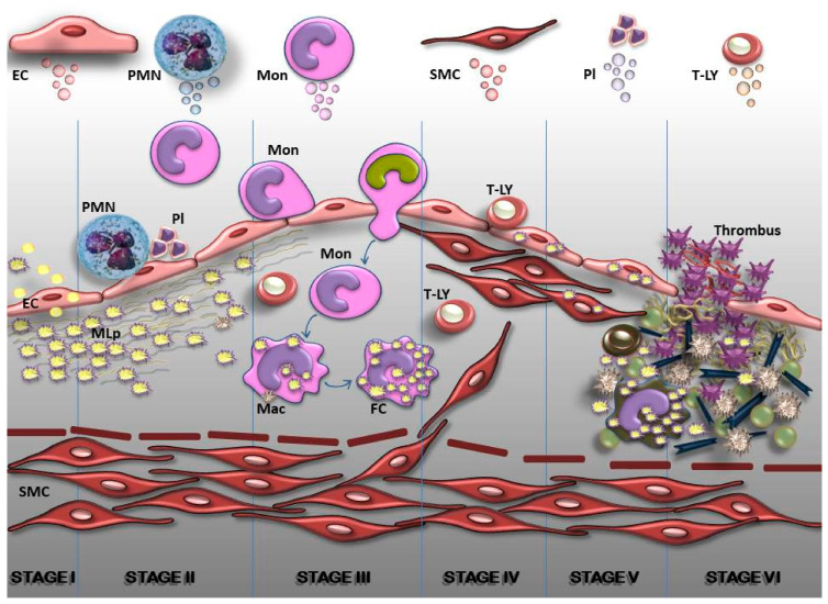
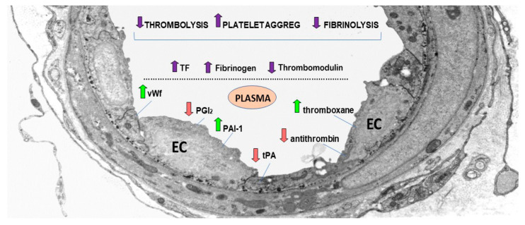
Cells convey information among one another. One instrument employed to transmit data and constituents to specific (target) cells is extracellular vesicles (EVs). They originate from a variety of cells (endothelial, immune cells, platelets, mesenchymal stromal cells, etc.), and consequently, their surface characteristics and cargo vary according to the paternal cell. The cargo could be DNA, mRNA, microRNA, receptors, metabolites, cytoplasmic proteins, or pathological molecules, as a function of which EVs exert different effects upon endocytosis in recipient cells. Recently, EVs have become important participants in a variety of pathologies, including atherogenesis and coronavirus disease 2019 (COVID-19)-associated thrombosis. Herein, we summarize recent advances and some of our own results on the role of EVs in atherosclerotic cardiovascular diseases, and discuss their potential to function as signaling mediators, biomarkers and therapeutic agents. Since COVID-19 patients have a high rate of thrombotic events, a special section of the review is dedicated to the mechanism of thrombosis and the possible therapeutic potential of EVs in COVID-19-related thrombosis. Yet, EV mechanisms and their role in the transfer of information between cells in normal and pathological conditions remain to be explored.

Keywords: COVID-19; atherosclerosis; cardiovascular disease; exosomes; extracellular vesicles; microvesicles; thrombosis.


Conflict of interest statement

The authors declare no conflict of interest.

Figures

Figure 1
Figure 1
Diagram representing the classification, biogenesis, molecular content and interactions with target cells of extracellular vesicle (EVs). Exosomes originate from the early sorting endosome (ESE), which turn sequentially into late sorting endosome (LSE), and multivesicular bodies (MVBs), whose intraluminal vesicles (ILVs) are exocytosed upon fusion and inward invagination of the plasmalemma. Ectosomes, also named microparticles (MPs) or microvesicles (MVs) are formed upon plasma membrane budding with phosphatidyl serine (PS) on the outer membrane surface. Apoptotic bodies (ApoBDs are membrane-derived large vesicles originating from apoptotic cells with PS as surface marker). (RME: receptor mediated endocytosis).
Figure 2
Figure 2
Diagram representing the arbitrary consecutive stages in the development of atherosclerotic plaques in arterial lesion-prone areas. Stage I. Endothelial cell (EC) activation/modulation of constitutive functions. The initial stage in atheroma formation is the accumulation within the plasma of oxidatively modified lipoproteins (MLp) that induce EC activation and increased transcytosis of MLp, i.e., their housing in the subendothelium where they interact with the components of the extracellular matrix (ECM). EC switch to a secretory phenotype that produces an excess of hyperplasic basal lamina. Stage II. EC dysfunction. Affected luminally by the alterations of plasma homoeostasis and abluminally by the accrual of MLp, the EC initiate an inflammatory process (i.e., the expression of new or more cell adhesion molecules, cytokines and chemokines) that attract immune cells such as neutrophils (PMN) which interact with platelets (Pl) and assist leucocyte migration. Stage III. Commencement of a robust inflammatory reaction. The recruitment of blood monocytes (Mon) and T lymphocytes (T-LY), diapedesis of Mon into the intima, where they become activated macrophages (Mac), expressing scavenger receptors which function in the uptake of MLp and the formation of foam cells (FC). Lymphocytes switch to activated pro-inflammatory (Th 1) and anti-inflammatory (Th 2 and TREG) cells that secrete cytokines and chemokines. Stage IV. SMC-key participants in the formation of fibrous plaques. The proliferation of resident SMC from the intima, and of SMC which has migrated from the media to the intima leads to the formation of a fibrous cap that is accompanied by increased synthesis of ECM. Stage V. Resident and immune cells and the factors they secrete generate a calcified fibro-lipid plaque. SMC, macrophages-derived foam cells, apoptotic cell-derived lipids and calcification centers form a necrotic core rich in cholesterol crystals. Stage VI. The unstable fibro-lipid plaque. Thinning of the fibrous cap, EC apoptosis and the accumulation of pro-inflammatory mediators leads to the physical rupture of the plaque. The direct contact between the ECM, the tissue factors and circulating platelets and blood coagulation components trigger thrombosis. In the upper part of the figure, the main cell sources of extracellular vesicles are shown.
Figure 3
Figure 3
Thrombosis is a consequence of reduced fibrinolysis and thrombolysis processes, and the subsequent dramatic increase in platelet aggregation due to an imbalance of endothelial and platelet circulating mediators. The figure shows an electron-micrograph of an arteriole exhibiting activated endothelial cells (EC), and the upregulated and downregulated mediators (arrows) that lead to thrombosis. An increase in plasma von Willebrand factor (vWF) that enhances platelet adhesivity, plasmin activator inhibitor (PAI-1) that inhibits tPA and uPA, and thromboxane A2 that activates the platelets, leads to platelet aggregation and the formation of thrombus. Concomitantly, thrombus formation is due to a decrease in plasma prostacyclin (PGI2) that controls platelet activation, tissue plasmin activator (tPA) that, together with urokinase plasminogen activator (uPA), breaks down fibrin into fibrin degradation products, and antithrombin (an active anticoagulant). Ultimately, the increase in tissue factor (TF) and fibrinogen, and the decrease of thrombomodulin, create a prothrombotic milieu by initiating the coagulation cascade, platelet aggregation and the formation of thrombus.

Similar articles

Cited by

References

    1. Boulanger C.M., Scoazec A., Ebrahimian T., Henry P., Mathieu E., Tedgui A., Mallat Z. Circulating Microparticles From Patients with Myocardial Infarction Cause Endothelial Dysfunction. Circulation. 2001;104:2649–2652. doi: 10.1161/hc4701.100516. - DOI - PubMed
    1. Boulanger C.M., Amabile N., Tedgui A. Circulating microparticles: A potential prognostic marker for atherosclerotic vascular disease. Hypertension. 2006;48:180–186. doi: 10.1161/01.HYP.0000231507.00962.b5. - DOI - PubMed
    1. Tokarz A., Szuścik I., Kuśnierz-Cabala B., Kapusta M., Konkolewska M., Żurakowski A., Georgescu A., Stępień E. Extracellular vesicles participate in the transport of cytokines and angiogenic factors in diabetic patients with ocular complications. Folia Med. Cracov. 2015;55:35–48. - PubMed
    1. Georgescu A., Alexandru N., Popov D., Amuzescu M., Andrei E., Zamfir C., Maniu H., Badila A. Chronic venous insufficiency is associated with elevated level of circulating microparticles. J. Thromb. Haemost. 2009;7:1566–1575. doi: 10.1111/j.1538-7836.2009.03525.x. - DOI - PubMed
    1. Georgescu A., Alexandru N., Andrei E., Titorencu I., Dragan E., Tarziu C., Ghiorghe S., Badila E., Bartos D., Popov D. Circulating microparticles and endothelial progenitor cells in atherosclerosis: Pharmacological effects of irbesartan. J. Thromb. Haemost. 2012;10:680–691. doi: 10.1111/j.1538-7836.2012.04650.x. - DOI - PubMed

MeSH terms