Review
. 2021 May 26;13(6):378.
doi: 10.3390/toxins13060378.

Therapeutic Uses of Bacterial Subunit Toxins

Affiliations
Review

Therapeutic Uses of Bacterial Subunit Toxins

Clifford Lingwood. Toxins (Basel). .

Abstract

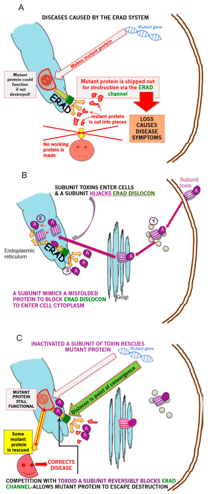
The B subunit pentamer verotoxin (VT aka Shiga toxin-Stx) binding to its cellular glycosphingolipid (GSL) receptor, globotriaosyl ceramide (Gb3) mediates internalization and the subsequent receptor mediated retrograde intracellular traffic of the AB5 subunit holotoxin to the endoplasmic reticulum. Subunit separation and cytosolic A subunit transit via the ER retrotranslocon as a misfolded protein mimic, then inhibits protein synthesis to kill cells, which can cause hemolytic uremic syndrome clinically. This represents one of the most studied systems of prokaryotic hijacking of eukaryotic biology. Similarly, the interaction of cholera AB5 toxin with its GSL receptor, GM1 ganglioside, is the key component of the gastrointestinal pathogenesis of cholera and follows the same retrograde transport pathway for A subunit cytosol access. Although both VT and CT are the cause of major pathology worldwide, the toxin-receptor interaction is itself being manipulated to generate new approaches to control, rather than cause, disease. This arena comprises two areas: anti neoplasia, and protein misfolding diseases. CT/CTB subunit immunomodulatory function and anti-cancer toxin immunoconjugates will not be considered here. In the verotoxin case, it is clear that Gb3 (and VT targeting) is upregulated in many human cancers and that there is a relationship between GSL expression and cancer drug resistance. While both verotoxin and cholera toxin similarly hijack the intracellular ERAD quality control system of nascent protein folding, the more widespread cell expression of GM1 makes cholera the toxin of choice as the means to more widely utilise ERAD targeting to ameliorate genetic diseases of protein misfolding. Gb3 is primarily expressed in human renal tissue. Glomerular endothelial cells are the primary VT target but Gb3 is expressed in other endothelial beds, notably brain endothelial cells which can mediate the encephalopathy primarily associated with VT2-producing E. coli infection. The Gb3 levels can be regulated by cytokines released during EHEC infection, which complicate pathogenesis. Significantly Gb3 is upregulated in the neovasculature of many tumours, irrespective of tumour Gb3 status. Gb3 is markedly increased in pancreatic, ovarian, breast, testicular, renal, astrocytic, gastric, colorectal, cervical, sarcoma and meningeal cancer relative to the normal tissue. VT has been shown to be effective in mouse xenograft models of renal, astrocytoma, ovarian, colorectal, meningioma, and breast cancer. These studies are herein reviewed. Both CT and VT (and several other bacterial toxins) access the cell cytosol via cell surface ->ER transport. Once in the ER they interface with the protein folding homeostatic quality control pathway of the cell -ERAD, (ER associated degradation), which ensures that only correctly folded nascent proteins are allowed to progress to their cellular destinations. Misfolded proteins are translocated through the ER membrane and degraded by cytosolic proteosome. VT and CT A subunits have a C terminal misfolded protein mimic sequence to hijack this transporter to enter the cytosol. This interface between exogenous toxin and genetically encoded endogenous mutant misfolded proteins, provides a new therapeutic basis for the treatment of such genetic diseases, e.g., Cystic fibrosis, Gaucher disease, Krabbe disease, Fabry disease, Tay-Sachs disease and many more. Studies showing the efficacy of this approach in animal models of such diseases are presented.

Keywords: endoplasmic reticulum associated degradation; neoplastic Gb3 expression; protein misfolding diseases; retrograde transport.


Conflict of interest statement

Lingwood is a founder of ERAD Therapeutics (eradtx.cpm) and has received travel support to present the new data reported herein.

Figures

Figure 1
Figure 1
Hijacking the hijacker. (A). Genetic diseases in which a small (e.g., point) mutation induces partial protein misfolding while retaining a significant fraction of the wildtype protein function, are candidates for this approach. The misfolded mutant is selected by ER chaperones for degradation by the cytosolic proteosome. Such proteins are unfolded, ubiquinylated and translocated across the ER membrane via the ERAD channel—the dislocon (indicated in green). This cytosolic destruction precipitates/exacerbates the deficiency disease symptoms. (B). Several bacterial subunit toxins hijack the ERAD translocon by A subunit mimicry of an unfolded protein. The holotoxin undergoes (GSL-dependent for VT and CT) retrograde from the cell surface to the ER. Subunit separation (via PDI) then allows the A subunit to enter and transit the dislocon which can only transport one polypeptide at a time. (C). Thus, the exogenous (inactivated toxin A subunit) and endogenous (mutant misfolded protein) translocation substrates converge at the ER dislocon. A subunit occupancy of the dislocon will compete for mutant protein transit, and thereby allow a fraction of the mutant protein to avoid ERAD and traffic to its correct cellular address to rescue (at least in part) the deficiency disease phenotype.
Figure 2
Figure 2
mCT restores F508delCFTR-dependent saliva production in the F508delCFTR mouse. Six Homozygous female F508delCFTR mice were injected i.v. with 400 ng (100 ng/mL blood) mCT every 48 h (from day 1) for two weeks. The dose was then increased to 1000 ng for a further three weeks, and CFTR dependent saliva secretion measured periodically (4 h post injection). Mice were left untreated for a further 11 days and saliva secretion measured. Three control mice were injected with saline and their saliva production did not change. No significant change in weight was observed for the treated mice, Saliva secretion in normal mice was measured as 6.7 µg/min/gm. After 40 days, the mice were left untreated for a further 10 days when the elevated saliva production returned to untreated levels. Importantly, no adverse effects were observed within the course of this study. These studies were performed under contract from ERAD Therapeutics by the Animal facilities at the Research Institute at the Hospital for Sick Children.
Figure 3
Figure 3
Effect of mCT on glucocerebrosidase A levels in N370 S Gaucher cells. N370S GBA Gaucher fibroblasts in 96 well microplate triplicates were treated with mCT as indicated and the expression level of GBA monitored by Western blot with MAB7410 using the quantitative Western Assay System (Protein Simple). These studies were carried out under contract by ERAD Therapeutics, in the laboratory of Dr Will Costain, NRC, Ottawa, ON, Canada. * p < 0.05, ** p < 0.01, *** p < 0.001.
Figure 4
Figure 4
mCT reduces plasma glucosyl sphingosine in the N370S Gaucher mouse. N370S glucocerebrosidase Gaucher mice (25 gm) were injected with 400 ng mCT18L i.p. every 48 h for two weeks. Blood samples taken prior to (day 0) and after (day 14) the treatment period were then assayed for glucosyl sphingosine by mass spectrometry and the prior and after values compared. Thus, each mouse served as its own control. The mouse studies were performed under contract from ERAD Therapeutics, in the laboratory of Dr Lorne Clark, UBC and the (blinded) blood glucosyl ceramide mass spectrometric analyses were performed at the facilities at the Hospital for Sick Children.

Similar articles

Cited by

References

    1. Karmali M., Petric M., Lim C., Fleming P.C., Arbus G.S., Lior H. The Association between Idiopathic Hemolytic Uremic Syndrome and Infection by Verotoxin-Producing Escherichia coli. J. Infect. Dis. 1985;151:775–782. doi: 10.1093/infdis/151.5.775. - DOI - PubMed
    1. Jokiranta T.S. HUS and atypical HUS. Blood. 2017;129:2847–2856. doi: 10.1182/blood-2016-11-709865. - DOI - PMC - PubMed
    1. Nakao H., Takeda T. Escherichia coli Shiga toxin. J. Nat. Toxins. 2000;9:299–313. - PubMed
    1. Ray P.E., Liu X.-H. Pathogenesis of Shiga toxin-induced hemolytic uremic syndrome. Pediatr. Nephrol. 2001;16:823–839. doi: 10.1007/s004670100660. - DOI - PubMed
    1. Khalid M., Andreoli S. Extrarenal manifestations of the hemolytic uremic syndrome associated with Shiga toxin-producing Escherichia coli (STEC HUS) Pediatr. Nephrol. 2019;34:2495–2507. doi: 10.1007/s00467-018-4105-1. - DOI - PubMed

Publication types

MeSH terms

LinkOut - more resources