

Potential Tamoxifen Repurposing to Combat Infections by Multidrug-Resistant Gram-Negative Bacilli
- PMID: 34073235
- PMCID: PMC8230278
- DOI: 10.3390/ph14060507
Potential Tamoxifen Repurposing to Combat Infections by Multidrug-Resistant Gram-Negative Bacilli
Abstract
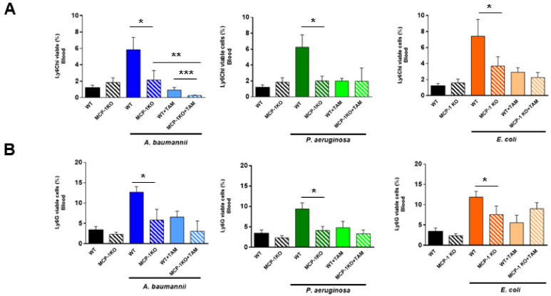
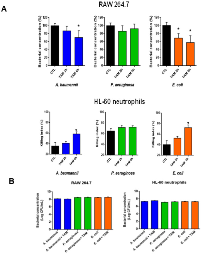
The development of new strategic therapies for multidrug-resistant bacteria, like the use of non-antimicrobial approaches and/or drugs repurposed to be used as monotherapies or in combination with clinically relevant antibiotics, has become urgent. A therapeutic alternative for infections by multidrug-resistant Gram-negative bacilli (MDR-GNB) is immune system modulation to improve the infection clearance. We showed that immunocompetent mice pretreated with tamoxifen at 80 mg/kg/d for three days and infected with Acinetobacter baumannii, Pseudomonas aeruginosa, or Escherichia coli in peritoneal sepsis models showed reduced release of the monocyte chemotactic protein-1 (MCP-1) and its signaling pathway interleukin-18 (IL-18), and phosphorylated extracellular signal-regulated kinase 1/2 (ERK1/2). This reduction of MCP-1 induced the reduction of migration of inflammatory monocytes and neutrophils from the bone marrow to the blood. Indeed, pretreatment with tamoxifen in murine peritoneal sepsis models reduced the bacterial load in tissues and blood, and increased mice survival from 0% to 60-100%. Together, these data show that tamoxifen presents therapeutic efficacy against MDR A. baumannii, P. aeruginosa, and E. coli in experimental models of infection and may be a new candidate to be repurposed as a treatment for GNB infections.
Keywords: animal model; bacteria; immune system; infection; repurposing drug; tamoxifen.
Conflict of interest statement
The authors declare no conflict of interest.
Figures


Similar articles
-
Repurposing of the Tamoxifen Metabolites to Combat Infections by Multidrug-Resistant Gram-Negative Bacilli.Antibiotics (Basel). 2021 Mar 22;10(3):336. doi: 10.3390/antibiotics10030336. Antibiotics (Basel). 2021. PMID: 33810067 Free PMC article.
-
Risk of acquiring multidrug-resistant Gram-negative bacilli from prior room occupants in the intensive care unit.Clin Microbiol Infect. 2011 Aug;17(8):1201-8. doi: 10.1111/j.1469-0691.2010.03420.x. Epub 2010 Dec 13. Clin Microbiol Infect. 2011. PMID: 21054665
-
Cefiderocol: A Siderophore Cephalosporin with Activity Against Carbapenem-Resistant and Multidrug-Resistant Gram-Negative Bacilli.Drugs. 2019 Feb;79(3):271-289. doi: 10.1007/s40265-019-1055-2. Drugs. 2019. PMID: 30712199 Review.
-
Temporal trends and patterns in antimicrobial-resistant Gram-negative bacteria implicated in intensive care unit-acquired infections: A cohort-based surveillance study in Istanbul, Turkey.J Glob Antimicrob Resist. 2018 Sep;14:190-196. doi: 10.1016/j.jgar.2018.04.015. Epub 2018 May 8. J Glob Antimicrob Resist. 2018. PMID: 29751127
-
ESCMID-EUCIC clinical guidelines on decolonization of multidrug-resistant Gram-negative bacteria carriers.Clin Microbiol Infect. 2019 Jul;25(7):807-817. doi: 10.1016/j.cmi.2019.01.005. Epub 2019 Jan 29. Clin Microbiol Infect. 2019. PMID: 30708122
Cited by
-
Repurposing of the Tamoxifen Metabolites to Combat Infections by Multidrug-Resistant Gram-Negative Bacilli.Antibiotics (Basel). 2021 Mar 22;10(3):336. doi: 10.3390/antibiotics10030336. Antibiotics (Basel). 2021. PMID: 33810067 Free PMC article.
-
Repurposing of the Tamoxifen Metabolites to Treat Methicillin-Resistant Staphylococcus epidermidis and Vancomycin-Resistant Enterococcus faecalis Infections.Microbiol Spectr. 2021 Oct 31;9(2):e0040321. doi: 10.1128/Spectrum.00403-21. Epub 2021 Oct 20. Microbiol Spectr. 2021. PMID: 34668743 Free PMC article.
-
Repurposing Tamoxifen as Potential Host-Directed Therapeutic for Tuberculosis.mBio. 2023 Feb 28;14(1):e0302422. doi: 10.1128/mbio.03024-22. Epub 2022 Dec 7. mBio. 2023. PMID: 36475748 Free PMC article.
-
Discovery of new antimicrobial thiophene derivatives with activity against drug-resistant Gram negative-bacteria.Front Pharmacol. 2024 Aug 20;15:1412797. doi: 10.3389/fphar.2024.1412797. eCollection 2024. Front Pharmacol. 2024. PMID: 39228527 Free PMC article.
-
Efficacy of Tamoxifen Metabolites in Combination with Colistin and Tigecycline in Experimental Murine Models of Escherichia coli and Acinetobacter baumannii.Antibiotics (Basel). 2024 Apr 24;13(5):386. doi: 10.3390/antibiotics13050386. Antibiotics (Basel). 2024. PMID: 38786115 Free PMC article.
References
-
- Garnacho-Montero J., Garcia-Garmendia J.L., Barrero-Almodovar A., Jiménez-Jiménez F.J., Pérez-Paredes C., Ortiz-Leyba C. Impact of adequate empirical antibiotic therapy on the outcome of patients admitted to the intensive care unit with sepsis. Crit. Care Med. 2003;31:2742–2751. doi: 10.1097/01.CCM.0000098031.24329.10. - DOI - PubMed
Grants and funding
LinkOut - more resources
Full Text Sources
Molecular Biology Databases
Research Materials
Miscellaneous
