Review
. 2021 May 26;10(11):2318.
doi: 10.3390/jcm10112318.

Old and New Challenges in Uveitis Associated with Behçet's Disease

Affiliations
Review

Old and New Challenges in Uveitis Associated with Behçet's Disease

Julie Gueudry et al. J Clin Med. .

Abstract

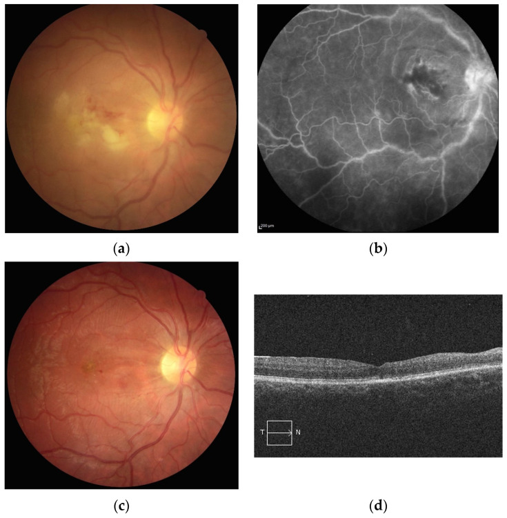
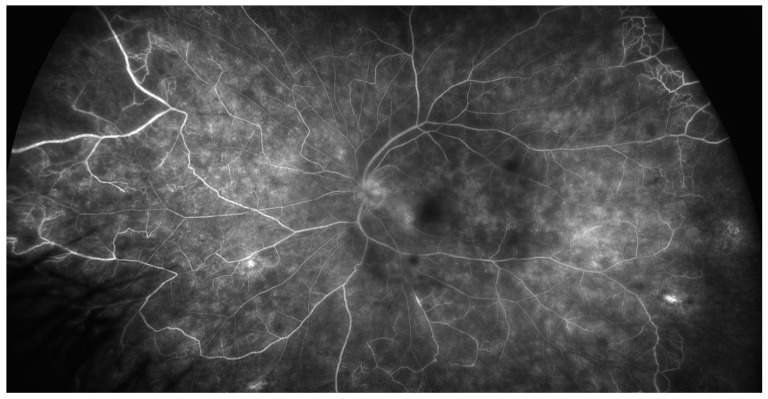
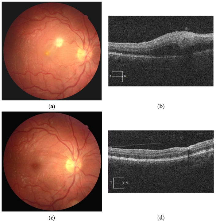
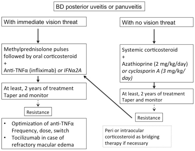
Behçet's disease (BD) is a systemic vasculitis disease of unknown origin occurring in young people, which can be venous, arterial or both, classically occlusive. Ocular involvement is particularly frequent and severe; vascular occlusion secondary to retinal vasculitis may lead to rapid and severe loss of vision. Biologics have transformed the management of intraocular inflammation. However, the diagnosis of BD is still a major challenge. In the absence of a reliable biological marker, diagnosis is based on clinical diagnostic criteria and may be delayed after the appearance of the onset sign. However, therapeutic management of BD needs to be introduced early in order to control inflammation, to preserve visual function and to limit irreversible structural damage. The aim of this review is to provide current data on how innovations in clinical evaluation, investigations and treatments were able to improve the prognosis of uveitis associated with BD.

Keywords: Behçet’s disease; anti-TNFα agent; biologics; retinal vasculitis; tocilizumab; uveitis.


Conflict of interest statement

The authors declare no conflict of interest.

Figures

Figure 1
Figure 1
(a) Retinal photography of the right eye of a patient with Behçet’s disease showing a white infiltrate in the inter-papillomacular area associated with retinal edema and hemorrhage. (b) Fluorescein angiography showing vascular staining in the involved area. (c) Two months later, retinal photography showing retinal and optic atrophy with resolution of the infiltrate. (d) Two months later, spectral domain OCT scan showing atrophy of the inner retinal layers.
Figure 2
Figure 2
Late phase of ultra-wide field fluorescein angiography showing papillary hyperfluorescence, extensive retinal capillaropathy and peripheral occlusive vasculitis in Behçet’s disease uveitis.
Figure 3
Figure 3
Late phase of ultra-wide field fluorescein angiography showing peripheral capillaropathy and vasculitis in the macular area responsible for visual acuity loss during Behçet’s disease uveitis.
Figure 4
Figure 4
(a) Retinal photography of the right eye showing area of whitish retinal necrosis related to Behçet’s disease and (b) its appearance on OCT as focal inner nuclear layer thickening; (c) its resolution 2 months later and (d) its appearance on OCT as focal inner nuclear layer collapse.
Figure 5
Figure 5
Proposed algorithm for management of severe Behçet’s disease (BD) uveitis.

Similar articles

  • Behçet's Disease Uveitis.
    Joubert M, Desbois AC, Domont F, Ghembaza A, Le Joncour A, Mirouse A, Maalouf G, Leclercq M, Touhami S, Cacoub P, Bodaghi B, Saadoun D. Joubert M, et al. J Clin Med. 2023 May 24;12(11):3648. doi: 10.3390/jcm12113648. J Clin Med. 2023. PMID: 37297843 Free PMC article. Review.
  • [Ocular manifestations in Behçet's disease].
    Desbois AC, Terrada C, Cacoub P, Bodaghi B, Saadoun D. Desbois AC, et al. Rev Med Interne. 2018 Sep;39(9):738-745. doi: 10.1016/j.revmed.2018.02.022. Epub 2018 Apr 4. Rev Med Interne. 2018. PMID: 29625716 Review. French.
  • Behçet's disease uveitis.
    Joubert M, Desbois AC, Domont F, Ghembaza A, Lejoncour A, Mirouse A, Maalouf G, Leclercq M, Touhami S, Cacoub P, Bodaghi B, Saadoun D. Joubert M, et al. Rev Med Interne. 2023 Oct;44(10):546-554. doi: 10.1016/j.revmed.2023.06.004. Epub 2023 Jul 4. Rev Med Interne. 2023. PMID: 37414674 Review.
  • Frosted branch angiitis and cerebral venous sinus thrombosis as an initial onset of neuro-Behçet's disease: a case report and review of the literature.
    Ferreira BFA, Rodriguez EEC, Prado LLD, Gonçalves CR, Hirata CE, Yamamoto JH. Ferreira BFA, et al. J Med Case Rep. 2017 Apr 15;11(1):104. doi: 10.1186/s13256-017-1261-z. J Med Case Rep. 2017. PMID: 28410605 Free PMC article. Review.
  • [Ocular manifestations in Behçet's disease].
    Zeghidi H, Saadoun D, Bodaghi B. Zeghidi H, et al. Rev Med Interne. 2014 Feb;35(2):97-102. doi: 10.1016/j.revmed.2013.10.011. Epub 2013 Nov 27. Rev Med Interne. 2014. PMID: 24290834 Review. French.

Cited by

References

    1. Evereklioglu C. Current concepts in the etiology and treatment of Behcet disease. Surv. Ophthalmol. 2005;50:297–350. doi: 10.1016/j.survophthal.2005.04.009. - DOI - PubMed
    1. Wechsler B., Du-Boutin L.T. Interferons and Behcet’s disease. Rev. Med. Interne. 2002;23:495s–499s. doi: 10.1016/S0248-8663(02)00665-3. - DOI - PubMed
    1. Cassoux N., Fardeau C., Lehoang P. Ocular manifestations of Behcet’s disease. Ann. Med. Interne. 1999;150:529–534. - PubMed
    1. Sakane T., Takeno M., Suzuki N., Inaba G. Behçet’s disease. N. Engl. J. Med. 1999;341:1284–1291. doi: 10.1056/NEJM199910213411707. - DOI - PubMed
    1. Mahr A., Maldini C. Epidemiology of Behçet’s disease. Rev. Med. Interne. 2014;35:81–89. doi: 10.1016/j.revmed.2013.12.005. - DOI - PubMed

LinkOut - more resources