Review
. 2021 May 26;13(6):792.
doi: 10.3390/pharmaceutics13060792.

Multifunctional Scaffolds and Synergistic Strategies in Tissue Engineering and Regenerative Medicine

Affiliations
Review

Multifunctional Scaffolds and Synergistic Strategies in Tissue Engineering and Regenerative Medicine

Nicolas Muzzio et al. Pharmaceutics. .

Abstract

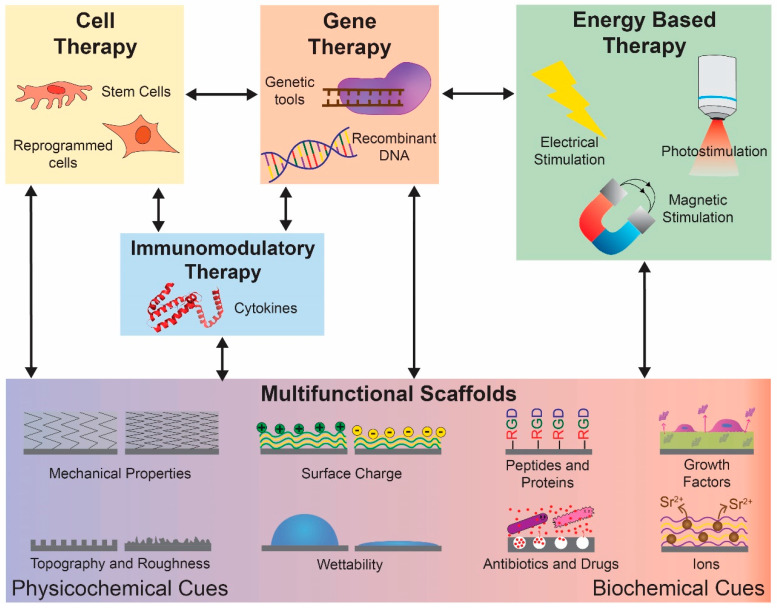
The increasing demand for organ replacements in a growing world with an aging population as well as the loss of tissues and organs due to congenital defects, trauma and diseases has resulted in rapidly evolving new approaches for tissue engineering and regenerative medicine (TERM). The extracellular matrix (ECM) is a crucial component in tissues and organs that surrounds and acts as a physical environment for cells. Thus, ECM has become a model guide for the design and fabrication of scaffolds and biomaterials in TERM. However, the fabrication of a tissue/organ replacement or its regeneration is a very complex process and often requires the combination of several strategies such as the development of scaffolds with multiple functionalities and the simultaneous delivery of growth factors, biochemical signals, cells, genes, immunomodulatory agents, and external stimuli. Although the development of multifunctional scaffolds and biomaterials is one of the most studied approaches for TERM, all these strategies can be combined among them to develop novel synergistic approaches for tissue regeneration. In this review we discuss recent advances in which multifunctional scaffolds alone or combined with other strategies have been employed for TERM purposes.

Keywords: biomaterials; combination therapy; multifunctional materials; scaffolds; tissue engineering and regenerative medicine (TERM).


Conflict of interest statement

The authors declare no conflict of interest.

Figures

Scheme 1
Scheme 1
Overview of different strategies and their combination with multifunctional scaffolds for tissue engineering and regenerative medicine.
Figure 1
Figure 1
Scaffolds combining synergistic physicochemical cues for TERM. (a) Schematic representation of anisotropic hydrogel preparation and cross-linking under the influence of a uniform magnetic field. Rod-shaped nanocrystals coated with MNPs, polydopamine and polyethylene glycol were employed to generate a gelatin hydrogel with directional microstructure and anisotropic mechanical properties. (b) Effect of isotropic and anisotropic hydrogels on human adipose tissue derived stem cells alignment after 3 days of culture (red, cytoskeleton; blue, nucleus). (c1,c2) SEM images of Ti implant and steam hydrothermal treated microarc oxidation coated Ti (ST-MAO), respectively. (d1,d2) Bone histology and histomorphometry around the Ti and ST-MAO implants, respectively, after 12 weeks of healing. (MB) mineralized bone; (black arrow) osteoblasts; (white arrow) osteocytes; (Coll) collagen birefringence; (yellow ring) osteon. (e1,e2) Field Emission-SEM TNTs and TNTs coated with PANI, respectively. (f) Antibacterial effect showing zone of inhibition on S. aureus. (a,b) adapted with permission from [133], American Chemical Society, 2019. (c1d2) adapted with permission from [134], American Chemical Society, 2015. (e1f) adapted with permission from [135], Elsevier, 2018.
Figure 2
Figure 2
Scaffolds combining synergistic biochemical cues for TERM. (a1,a2) Gross morphology and microstructure of Li- and Si-containing scaffolds. (b1c2) In vivo osteochondral regeneration efficiency for Li- and Si-containing scaffolds 12 weeks postsurgery: macrophotograph (b1,b2) and transverse view of 3D reconstruction of microcomputed tomography (c1,c2) showing the defects in control (untreated) (b1,c1) and implanted with scaffold (b2,c2). In microcomputed tomography the off-white color, green color and red color stand for primary bone, new bone, and scaffold, respectively. (d1,d2) SEM morphologies of untreated and after degradation for 4 weeks of PCL/polysialic acid/methylprednisolone nanofiber scaffolds, respectively. (e) Photographs of spinal tissue, spinal cord transection and spinal cord after nanofiber transplantation. (f) Histology and quantification of myelin sheaths 7 weeks postoperation. The dotted lines indicate the plane of injury. The letter R and C represents rostral and caudal spinal cord. SCI: spinal cord injury. (a1c2) adapted with permission from [140], Elsevier, 2017. (d1f) adapted with permission from [141], Elsevier, 2018.
Figure 3
Figure 3
Scaffold combining synergistic physicochemical and biochemical cues for TERM. (a) Schematic representation of the fabrication of porous HA scaffolds covered with a BG-containing PLGA fiber layer (HPB). (b1,b2) Representative SEM images of porous HA and HPB scaffolds, respectively. The thickness of BG-containing PLGA microfibers is shown. (c1,c2) Osteogenic activity of MC3T3-E1 cells cultured on HA and HPB scaffolds, respectively. Immunocytochemical analyses of the osteogenic protein expression level of Col I (COL-I, green), runt-related transcription factor 2 (RUNX2, red) and osteopontin (OPN, magenta) following culture for 14 days. (d1,d2) Alizarin red staining performed to observe mineralization of MC3T3-E1 cells by ions released around HA and HPB scaffolds, respectively, on day 21. (ad2) adapted with permission from [154], American Chemical Society, 2019. Further permissions to the related material should be directed to the ACS.
Figure 4
Figure 4
Synergistic approaches combining multifunctional scaffolds with cell-based therapy for TERM. (a) Representative optical images of native and decellularized heart tissue. (b) Masson’s trichrome staining confirming absence of cells and cell debris in the matrix after decellularization. (c) Gross appearance of the volumetric muscle loss injury in four groups and (d) surface ratio of new muscle fibers formed in each group. MSCs and ECM scaffold have a synergistic effect promoting muscle tissue regeneration. Data shown as mean ± SD. ** p < 0.01; *** p < 0.001; NS, not significant (p > 0.05). PBS phosphate buffer saline. (e) Scheme of the adhesive peptide modified hyaluronic acid scaffold loaded with bone marrow MSCs for spinal cord injury repair. (f) H&E staining of nontreated tissues (spinal cord injury) and tissues embedded with implants at 4 weeks postsurgery. Encapsulation of MSCs significantly improved nerve tissue reconnection effect of the scaffold. (ad) adapted from [159], BioMed Central, 2018. (ef) adapted with permission from [161], American Chemical Society, 2017.
Figure 5
Figure 5
Synergistic approaches combining multifunctional scaffolds with gene or immune therapy for TERM. (a) Schematic diagram of scaffold fabrication through coating gelatin-conjugated caffeic acid (GelCA) onto the surface of apatite-PLGA (Ap-PLGA) scaffold. The GelCA-coated hybrid biopolymer scaffold was adopted to deliver adeno-associated viruses-encoding Trb3 or/and recombinant BMP-2 protein. (bd) Promotion of bone formation (b,c) and fat-filled cyst formation inhibition in rat critical-sized mandibular defects 12 weeks after scaffold implantation loaded with high dose BMP-2 (hBMP2) and/or low and high dose adeno-associated viruses-encoding Trb3 (Trb3-lv and Trb3-hv, respectively). (b) Bone volume/tissue volume percentage. (c) Bone mineral density. Values represent mean ± SD: ** p < 0.01 and *** p < 0.001 using one-way ANOVA test. (d) Oil red immunohistochemical stain showing fewer adipocyte-like cells in area of defects treated with high dose AAV-Trb3 and BMP-2 than with BMP-2 alone. (e) Transgene expression in vivo. Representative bioluminescence images at days 3, 7, and 28 postimplantation of scaffolds loaded with luciferase lentivirus and implanted into the right intraperitoneal fat pad. Color bar indicates radiance (p/sec/cm2/sr). (f) Leukocyte infiltration into IL-10 virus releasing scaffolds. Number of CD45 positive cells isolated from scaffolds loaded with IL-10 or luciferase virus at day 3 and day 7 post implantation. * p < 0.05 versus day 7 luciferase. (ad) adapted with permission from [164], Elsevier, 2020. (ef) adapted with permission from [165], Elsevier, 2013.
Figure 6
Figure 6
Synergistic approaches combining multifunctional scaffolds with energy-based therapy for TERM. (a) Schematic illustration of the fabrication of nHA/GO particles, nHA/GO/chitosan scaffolds, and their bioapplication. (b1,b2) Representative tumor photographs of control group (without adding any scaffolds or irradiation) and nHA/GO/chitosan scaffold implanted and daily irradiated tumors, respectively. (c1d2) H&E and Masson histological analysis of the new bone formation (black arrows, new bone; green arrows, collagen fiber). (c1,d1) control group. (c2,d2) irradiated nHA/GO/chitosan scaffolds. (e) Construction of a smart electroactive tissue engineering scaffold with ability to control release and expression of BMP-4 for efficient bone repair. (f1,f2) In vivo repair of smart electroactive polymer scaffolds. X-ray detection of rabbit radial defect implanted with PLGA/HA scaffold without electrical stimulation (f1) and PLGA/HA/PLA-aniline pentamer/BMP-4 scaffold with electrical stimulation (f2). Four images from top to bottom were collected in 2-, 4-, 8-, or 12-weeks postoperation for each group. (ad2) adapted with permission from [177], Elsevier, 2019. (ef2) adapted with permission from [179], Elsevier, 2019.

Similar articles

Cited by

References

    1. Qu H., Fu H., Han Z., Sun Y. Biomaterials for bone tissue engineering scaffolds: A review. RSC Adv. 2019;9:26252–26262. doi: 10.1039/C9RA05214C. - DOI - PMC - PubMed
    1. Han F., Wang J., Ding L., Hu Y., Li W., Yuan Z., Guo Q., Zhu C., Yu L., Wang H., et al. Tissue Engineering and Regenerative Medicine: Achievements, Future, and Sustainability in Asia. Front. Bioeng. Biotechnol. 2020;8 doi: 10.3389/fbioe.2020.00083. - DOI - PMC - PubMed
    1. Frey B.M., Zeisberger S.M., Hoerstrup S.P. Tissue Engineering and Regenerative Medicine-New Initiatives for Individual Treatment Offers. Transfus. Med. Hemotherapy. 2016;43:318–320. doi: 10.1159/000450716. - DOI - PMC - PubMed
    1. Dzobo K., Thomford N.E., Senthebane D.A., Shipanga H., Rowe A., Dandara C., Pillay M., Motaung K.S.C.M. Advances in Regenerative Medicine and Tissue Engineering: Innovation and Transformation of Medicine. Stem Cells Int. 2018;2018:2495848. doi: 10.1155/2018/2495848. - DOI - PMC - PubMed
    1. Walma D.A.C., Yamada K.M. The extracellular matrix in development. Development. 2020;147:dev175596. doi: 10.1242/dev.175596. - DOI - PMC - PubMed

LinkOut - more resources