

Camu-Camu Fruit Extract Inhibits Oxidative Stress and Inflammatory Responses by Regulating NFAT and Nrf2 Signaling Pathways in High Glucose-Induced Human Keratinocytes
- PMID: 34073317
- PMCID: PMC8198278
- DOI: 10.3390/molecules26113174
Camu-Camu Fruit Extract Inhibits Oxidative Stress and Inflammatory Responses by Regulating NFAT and Nrf2 Signaling Pathways in High Glucose-Induced Human Keratinocytes
Abstract
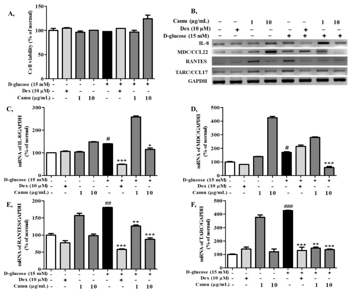
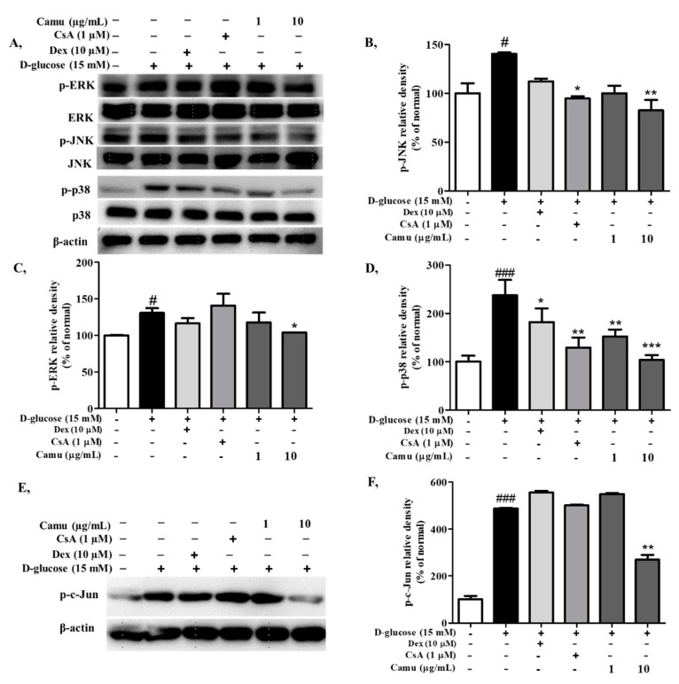
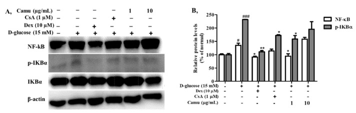
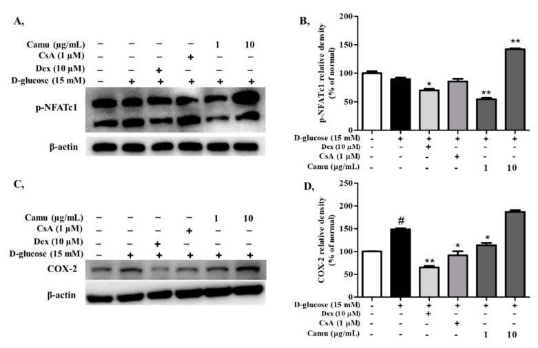
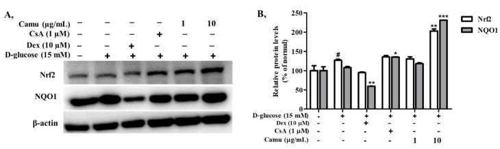
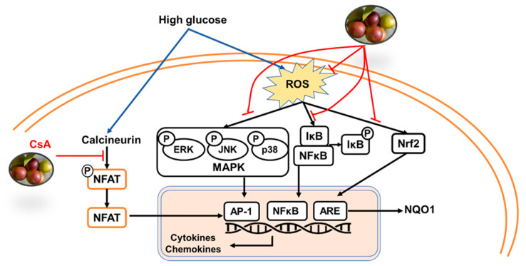
Myrciaria dubia (HBK) McVaugh (camu-camu) belongs to the family Myrtaceae. Although camu-camu has received a great deal of attention for its potential pharmacological activities, there is little information on the anti-oxidative stress and anti-inflammatory effects of camu-camu fruit in skin diseases. In the present study, we investigated the preventative effect of 70% ethanol camu-camu fruit extract against high glucose-induced human keratinocytes. High glucose-induced overproduction of reactive oxygen species (ROS) was inhibited by camu-camu fruit treatment. In response to ROS reduction, camu-camu fruit modulated the mitogen-activated protein kinases (MAPK)/activator protein-1 (AP-1), nuclear factor kappa-light-chain-enhancer of activated B cells (NF-κB), and nuclear factor of activated T cells (NFAT) signaling pathways related to inflammation by downregulating the expression of proinflammatory cytokines and chemokines. Furthermore, camu-camu fruit treatment activated the expression of nuclear factor E2-related factor 2 (Nrf2) and subsequently increased the NAD(P)H:quinone oxidoreductase1 (NQO1) expression to protect keratinocytes against high-glucose-induced oxidative stress. These results indicate that camu-camu fruit is a promising material for preventing oxidative stress and skin inflammation induced by high glucose level.
Keywords: NFAT; Nrf2; camu-camu fruit; high glucose; inflammation; keratinocytes; oxidative stress.
Conflict of interest statement
The authors declare no conflict of interest.
Figures

Similar articles
-
Anti-Allergic Effects of Myrciaria dubia (Camu-Camu) Fruit Extract by Inhibiting Histamine H1 and H4 Receptors and Histidine Decarboxylase in RBL-2H3 Cells.Antioxidants (Basel). 2021 Dec 31;11(1):104. doi: 10.3390/antiox11010104. Antioxidants (Basel). 2021. PMID: 35052608 Free PMC article.
-
Crataegus laevigata Suppresses LPS-Induced Oxidative Stress during Inflammatory Response in Human Keratinocytes by Regulating the MAPKs/AP-1, NFκB, and NFAT Signaling Pathways.Molecules. 2021 Feb 6;26(4):869. doi: 10.3390/molecules26040869. Molecules. 2021. PMID: 33562140 Free PMC article.
-
Nrf2 deficiency induces oxidative stress and promotes RANKL-induced osteoclast differentiation.Free Radic Biol Med. 2013 Dec;65:789-799. doi: 10.1016/j.freeradbiomed.2013.08.005. Epub 2013 Aug 14. Free Radic Biol Med. 2013. PMID: 23954472
-
Camu-camu [Myrciaria dubia (HBK) McVaugh]: A review of properties and proposals of products for integral valorization of raw material.Food Chem. 2022 Mar 15;372:131290. doi: 10.1016/j.foodchem.2021.131290. Epub 2021 Oct 2. Food Chem. 2022. PMID: 34818735 Review.
-
Activation of Nrf2/HO-1 signaling: An important molecular mechanism of herbal medicine in the treatment of atherosclerosis via the protection of vascular endothelial cells from oxidative stress.J Adv Res. 2021 Jul 6;34:43-63. doi: 10.1016/j.jare.2021.06.023. eCollection 2021 Dec. J Adv Res. 2021. PMID: 35024180 Free PMC article. Review.
Cited by
-
Feasibility of Catalpol Intranasal Administration and Its Protective Effect on Acute Cerebral Ischemia in Rats via Anti-Oxidative and Anti-Apoptotic Mechanisms.Drug Des Devel Ther. 2022 Jan 25;16:279-296. doi: 10.2147/DDDT.S343928. eCollection 2022. Drug Des Devel Ther. 2022. PMID: 35115763 Free PMC article.
-
Anti-Allergic Effects of Myrciaria dubia (Camu-Camu) Fruit Extract by Inhibiting Histamine H1 and H4 Receptors and Histidine Decarboxylase in RBL-2H3 Cells.Antioxidants (Basel). 2021 Dec 31;11(1):104. doi: 10.3390/antiox11010104. Antioxidants (Basel). 2021. PMID: 35052608 Free PMC article.
-
Nutritional Composition, Pharmacological Properties, and Industrial Applications of Myrciaria dubia: An Undiscovered Superfruit.Food Sci Nutr. 2025 May 23;13(6):e70331. doi: 10.1002/fsn3.70331. eCollection 2025 Jun. Food Sci Nutr. 2025. PMID: 40417739 Free PMC article. Review.
-
Two Cases of Durable and Deep Responses to Immune Checkpoint Inhibition-Refractory Metastatic Melanoma after Addition of Camu Camu Prebiotic.Curr Oncol. 2023 Aug 25;30(9):7852-7859. doi: 10.3390/curroncol30090570. Curr Oncol. 2023. PMID: 37754485 Free PMC article.
-
Targeted activation of Nrf2/HO-1 pathway by Corynoline alleviates osteoporosis development.Food Sci Nutr. 2023 Jan 24;11(4):2036-2048. doi: 10.1002/fsn3.3239. eCollection 2023 Apr. Food Sci Nutr. 2023. PMID: 37051369 Free PMC article.
References
-
- Rufino M., Socorro M.D., Alves R.E., de Brito E.S., Pérez-Jiménez J., Saura-Calixto F., Mancini-Filho J. Bioactive Compounds and Antioxidant Capacities of 18 Non-Traditional Tropical Fruits from Brazil. Food Chem. 2010;121:996–1002. doi: 10.1016/j.foodchem.2010.01.037. - DOI
-
- Castro J.C., Maddox J.D., Imán S.A. Exotic Fruits. Elsevier; Amsterdam, The Netherlands: 2018. Camu-camu—Myrciaria dubia (Kunth) McVaugh; pp. 97–105.
-
- Azevêdo J.C.S., Borges K.C., Genovese M.I., Correia R.T.P., Vattem D.A. Neuroprotective Effects of Dried Camu-Camu (Myrciaria dubia HBK McVaugh) Residue in C. Elegans. Food Res. Int. 2015;73:135–141. doi: 10.1016/j.foodres.2015.02.015. - DOI
-
- Fujita A., Sarkar D., Wu S., Kennelly E., Shetty K., Genovese M.I. Evaluation of Phenolic-Linked Bioactives of Camu-Camu (Myrciaria dubia Mc. Vaugh) for Antihyperglycemia, Antihypertension, Antimicrobial Properties and Cellular Rejuvenation. Food Res. Int. 2015;77:194–203. doi: 10.1016/j.foodres.2015.07.009. - DOI
MeSH terms
Substances
LinkOut - more resources
Full Text Sources
Research Materials
Miscellaneous
