. 2021 May 26;22(11):5650.
doi: 10.3390/ijms22115650.

In Vitro Framework to Assess the Anti- Helicobacter pylori Potential of Lactic Acid Bacteria Secretions as Alternatives to Antibiotics

Affiliations

In Vitro Framework to Assess the Anti- Helicobacter pylori Potential of Lactic Acid Bacteria Secretions as Alternatives to Antibiotics

Samantha A Whiteside et al. Int J Mol Sci. .

Abstract

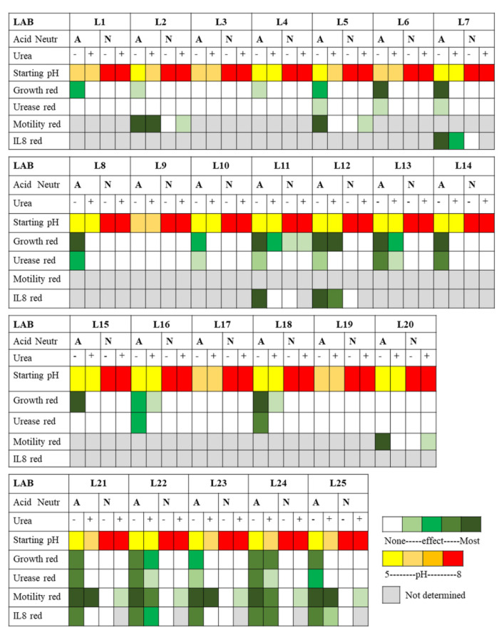
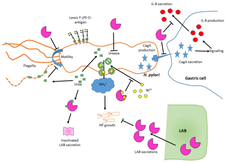
Helicobacter pylori is a prevalent bacterium that can cause gastric ulcers and cancers. Lactic acid bacteria (LAB) ameliorate treatment outcomes against H. pylori, suggesting that they could be a source of bioactive molecules usable as alternatives to current antibiotics for which resistance is mounting. We developed an in vitro framework to compare the anti-H. pylori properties of 25 LAB and their secretions against H. pylori. All studies were done at acidic and neutralized pH, with or without urea to mimic various gastric compartments. Eighteen LAB strains secreted molecules that curtailed the growth of H. pylori and the activity was urea-resistant in five LAB. Several LAB supernatants also reduced the urease activity of H. pylori. Pre-treatment of H. pylori with acidic LAB supernatants abrogated its flagella-mediated motility and decreased its ability to elicit pro-inflammatory IL-8 cytokine from human gastric cells, without reverting the H. pylori-induced repression of other pro-inflammatory cytokines. This study identified the LAB that have the most anti-H. pylori effects, decreasing its viability, its production of virulence factors, its motility and/or its ability to elicit pro-inflammatory IL-8 from gastric cells. Once identified, these molecules can be used as alternatives or complements to current antibiotics to fight H. pylori infections.

Keywords: Helicobacter pylori; cytokines; gastric disease; inflammation; lactic acid bacteria; secretome; urease.


Conflict of interest statement

The authors C.C., S.A.W., M.M.M., S.S. and J.J. declare no conflict of interest. The authors T.A.T. and C.W.M. are LHS employees who participated to this study and LHS funded part of these studies.

Figures

Figure 1
Figure 1
Anti-H. pylori activity of LAB in overlay assays. The LAB were grown embedded in the bottom layer (CBA or mMRS) of bi-layered plates. The top layer was always CBA. Afterwards, H. pylori was applied to the top layer and its growth was monitored after two days of incubation by harvesting the bacterial lawn and measuring the OD600nm of the suspension. The data were normalized to the OD600nm of the negative control that did not comprise any LAB; n = 2 to 5 biological replicates, with 1 technical replicate per biological replicate. Significance was assessed by one-way ANOVA with Dunnett’s multiple comparison test using the control without H. pylori and prepared in the same media as comparator; a: p < 0.05; c: p < 0.001; d: p < 0.0001; ns: not significant.
Figure 2
Figure 2
Effect of L21–L25 LAB supernatants on the growth and urease activity of H. pylori NCTC 11637. LAB supernatants from strains L21 to L25 were harvested after 12 h or 24 h of growth in mMRS broth under anaerobic conditions. H. pylori (HP) strain NCTC 11637 was exposed to 25% of these supernatants or control media without urea (hatched bars) or with 10 mM urea (solid bars) for 48 h. Growth was determined by spot plating and counting colony forming units (CFU) (A). The lower limit of detection for the assay was 103 CFU/mL. The H. pylori cultures were also tested for the intracellular urease activity using the Berthelot reaction (B). The urease data are represented as the log2 fold change of the NH4+ produced by 180 µL of resuspended bacterial pellet over 30 min relative to the urea-free untreated control (BHI). The legend indicated in (B) applies to both panels. Specifically, the supernatants were left unmodified at their acidic pH (denoted A, blue and orange bars) or were neutralized to pH 7 by NaOH (denoted N, green and yellow bars). Controls included LAB growth media (LM), LM acidified to pH 4.5 by HCl (HCl), acidified to pH 4.5 by lactic acid (LA), or acidified to pH 4.5 by lactic acid then neutralized with NaOH (LN). As a positive control, H. pylori cultured in BHI without LAB supernatant or media were included. For each LAB, threeto five biological replicates comprising each threetechnical replicates were performed. For the controls, fiveto eightbiological replicates were performed. Significance was assessed by two-way ANOVA with Sidak multiple comparison test. BHI with matching time point and matching urea concentration was used as the comparator for each data set. Significance was represented as a, p < 0.05; b, p < 0.01; c, p < 0.001; and d, p < 0.0001. Lack of significance label indicates no significance.
Figure 3
Figure 3
Effects of exposure to LAB L1–L20 supernatants on the viability and urease activity of H. pylori NCTC 11637. All LAB supernatants were harvested after 24 h of growth and H. pylori growth (A) and intracellular urease activity (B) were measured as described in Figure 2. Significance was represented as a, p < 0.05; b, p < 0.01; and d, p < 0.0001. Lack of significance label indicates no significance. For (A), three to four biological replicates were performed for each strain and ten to fifteen replicates of controls. For (B), four to five biological replicates were performed for each strain and eleven to fifteen replicates of controls.
Figure 4
Figure 4
The activity of several LAB is due to bioactive molecules that exert their effects beyond pH effects. Exposure assays were carried out with media acidified with HCl in varying concentrations to generate a pH standard curve (A,B) or with LAB supernatants (C). The pH was monitored immediately after mixing each acidified media or LAB supernatant with H. pylori or after 48 h of exposure. The effect of HCl-mediated acidity on H. pylori recovery was recorded after 48 h of incubation (B). The effects of the LAB on H. pylori recovery were as presented in Figure 2 and Figure 3. The legend box applies to all panels. Specifically, samples with starting acidic pH are represented in orange (0 h) and blue (48 h) while samples with original neutral pH are represented in yellow (0 h) and green (48 h). Hatched bars signify the lack of urease while solid bars signify the presence of urea. Statistics were performed by one-way ANOVA with Dunnett’s multiple comparison test, using BHI of matching time and urea concentration as comparator. Significance was represented as a, p < 0.05; b, p < 0.01; c, p < 0.001; and d, p < 0.0001. Lack of significance label indicates no significance. For (A,B), two to three biological replicates were performed. For (C), three biological replicates were performed for each LAB strain, and four biological replicates were performed for the controls.
Figure 5
Figure 5
Effects of LAB supernatants and pH on H. pylori motility. H. pylori was stabbed in soft agar after 48 h of exposure to LAB or control, in the presence (solid bar) or absence (hatched bars) of urea. Samples with starting acidic pH (denoted A) are represented in blue while samples with starting neutral pH (denoted N) are represented in green. nd: none detected. Statistics performed as per Figure 2 relative to the BHI/no urea control. Statistics were performed by one-way ANOVA with Dunnett’s multiple comparison test, using BHI with matching urea concentration as comparator. Black letters for “BHI no urea” comparator and blue letters for “BHI + urea” comparator. Significance was represented as c, p < 0.001; and d, p < 0.0001 with two to three biological replicates per sample. Lack of significance label indicates no significance.
Figure 6
Figure 6
Effect of exposure of H. pylori to LAB supernatants on H. pylori-mediated secretion of IL-8 by gastric cells. IL-8 was measured in the supernatant of AGS cells exposed to H. pylori NCTC 11637 that had been pre-exposed to LAB supernatants or control media. The supernatants were left unmodified (acidic, A, blue bars) and added to H. pylori cultures without urea (hatched bars) or with 10 mM urea (solid bars); or were neutralized to pH 7 by NaOH (N, green bars) and added to H. pylori cultures without urea (hatched bars). Neutralized samples were not tested with urea, thus “nd” here indicates “not determined”. Controls included LAB growth media (LM), LM acidified to pH 4.5 by HCl (HCl), acidified to pH 4.5 by lactic acid (LA), or acidified to pH 4.5 by lactic acid then neutralized with NaOH (LN). As a positive control, H. pylori cultured in BHI without LAB supernatant or media control were included. All H. pylori controls were also completed in the presence or absence of 10 mM urea and followed the same color coding as test samples. Untreated AGS cell supernatant was included as a baseline (No HP). The data were derived from multiplex (27- and 40-plex Luminex assays) and ELISA experiments performed on the same samples, with three biological replicates in each experiment. The 27-plex and the ELISA data were normalized to the 40-plex data using the ratio of the average “urea-free, BHI” control values relative to the average “urea-free, BHI” control value of the 40-plex data. Statistics were performed by one-way ANOVA with Dunnett’s multiple comparison test. Significance was represented as b, p < 0.01; c, p < 0.001; and d, p < 0.0001. Lack of statistical label indicates no significance. Urea-free LA and LA containing 10 mM urea are the comparators for all acidic supernatants in the presence and absence of 10 mM of urea, depicted respectively in black and blue letters. Urea-free LN is the comparator for the pH adjusted (neutralized) supernatants in the absence of urea (no significance letters as none were significant). In addition, the “No HP” sample was used as a comparator to all test samples to demonstrate activation or lack thereof upon addition of H. pylori (red letters).
Figure 7
Figure 7
Effects of exposure of H. pylori to L25 supernatants on H. pylori-mediated secretion of cytokines by gastric cells. The data shown are extracted from Figure 6 and Figure S4 for NCTC 11637, with three biological replicates per sample. Only cytokines for which most differences were observed are shown, as well as 3 cytokines for which no effect occurred to demonstrate the specificity of the phenotypes. The supernatant left at its acidic pH (L25A) is compared to the LA media (hatched blue bars without urea; solid blue bars with 10 mM urea). The supernatant that was neutralized (L25N) is compared to the LN control media (hatched green bar, only determined in absence of urea). As a positive control, H. pylori cultured in BHI without LAB supernatant or media control were included (hatched black bars). Statistics were performed by 1-way ANOVA with Dunnett’s multiple comparison test. Significance was represented as a, p < 0.05 and b, p < 0.01. Urea-free LA and LA containing 10 mM urea are the comparators for all acidic supernatants in the presence and absence of 10 mM of urea, respectively. Urea-free LN is the comparator for the pH adjusted (neutralized) supernatants in the absence of urea. Lack of statistical label indicates no significance.
Figure 8
Figure 8
Heat map summary of all effects measured for the 25 LAB supernatants on H. pylori NCTC 11637. The supernatants were harvested at 24 h and used as their native acidic pH (A) or neutralized to pH 7 (N, Neutr). The starting pH of the exposure mixture is color coded from yellow (pH 4.5) to red (pH 8). For phenotypic effects, the color coding is relative to non-exposed H. pylori and depicts the reduction (red) of the original feature, from white for no effect to dark green for most effects. Grey indicates that no determination was performed.
Figure 9
Figure 9
Model highlighting the various effects of LAB secretions on H. pylori virulence features. Activities vary across LAB and encompass direct effects on H. pylori growth, reduction of intracellular urease activity probably due to nickel chelation, abrogation of H. pylori’s ability to elicit IL-8 production likely via impairment of CagA secretion, and/or abrogation of motility. No effect was seen on flagellin, CagA or Lewis O-antigen production. Urea was able to inactivate some LAB secretions and to decrease H. pylori motility, likely via its chaotropic nature.

Similar articles

Cited by

References

    1. Marshall B.J., Warren J.R. Unidentified Curved Bacilli in The Stomach of Patients with Gastritis and Peptic Ulceration. Lancet. 1984;323:1311–1315. doi: 10.1016/S0140-6736(84)91816-6. - DOI - PubMed
    1. Wang F., Meng W., Wang B., Qiao L. Helicobacter pylori-induced gastric inflammation and gastric cancer. Cancer Lett. 2014;345:196–202. doi: 10.1016/j.canlet.2013.08.016. - DOI - PubMed
    1. Fox J.G., Wang T.C. Inflammation, atrophy, and gastric cancer. J. Clin. Investig. 2007;117:60–69. doi: 10.1172/JCI30111. - DOI - PMC - PubMed
    1. Malfertheiner P., Sipponen P., Naumann M., Moayyedi P., Mégraud F., Xiao S.-D., Sugano K., Nyrén O., Force T.L.H.P.-G.C.T. Helicobacter pylori Eradication Has the Potential to Prevent Gastric Cancer: A State-of-the-Art Critique. Am. J. Gastroenterol. 2005;100:2100–2115. doi: 10.1111/j.1572-0241.2005.41688.x. - DOI - PubMed
    1. Ekström A.M., Held M., Hansson L.-E., Engstrand L., Nyrén O. Helicobacter pylori in gastric cancer established by CagA immunoblot as a marker of past infection. Gastroenterol. 2001;121:784–791. doi: 10.1053/gast.2001.27999. - DOI - PubMed

LinkOut - more resources