Review
. 2021 May 26;10(6):1325.
doi: 10.3390/cells10061325.

EFA6 in Axon Regeneration, as a Microtubule Regulator and as a Guanine Nucleotide Exchange Factor

Affiliations
Review

EFA6 in Axon Regeneration, as a Microtubule Regulator and as a Guanine Nucleotide Exchange Factor

Gilberto Gonzalez et al. Cells. .

Retraction in

Abstract

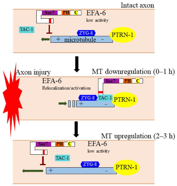
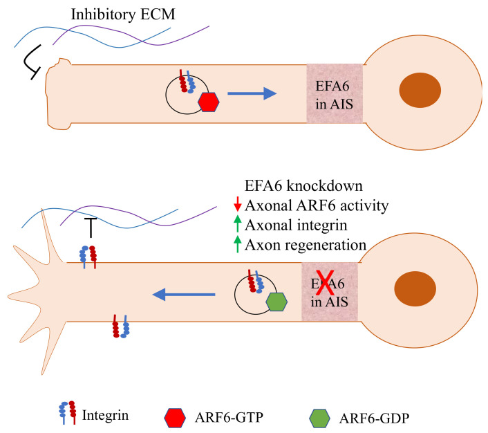
Axon regeneration after injury is a conserved biological process that involves a large number of molecular pathways, including rapid calcium influx at injury sites, retrograde injury signaling, epigenetic transition, transcriptional reprogramming, polarized transport, and cytoskeleton reorganization. Despite the numerous efforts devoted to understanding the underlying cellular and molecular mechanisms of axon regeneration, the search continues for effective target molecules for improving axon regeneration. Although there have been significant historical efforts towards characterizing pro-regenerative factors involved in axon regeneration, the pursuit of intrinsic inhibitors is relatively recent. EFA6 (exchange factor for ARF6) has been demonstrated to inhibit axon regeneration in different organisms. EFA6 inhibition could be a promising therapeutic strategy to promote axon regeneration and functional recovery after axon injury. This review summarizes the inhibitory role on axon regeneration through regulating microtubule dynamics and through affecting ARF6 (ADP-ribosylation factor 6) GTPase-mediated integrin transport.

Keywords: ARF6; EFA6; axon injury; axon regeneration; integrin; microtubule dynamics.


Conflict of interest statement

The authors declare no conflict of interest.

Figures

Figure 1
Figure 1
The conserved EFA6 protein family. (A) Graphic illustration of EFA6 family proteins showing the PH (Pleckstrin Homology), Sec7 (GEF activity) and CC (coiled coil) domains, based on sequences from NCBI. Not to scale. An 18 a.a. motif (microtubule elimination domain, MTED) is present in C. elegans and Drosophila EFA6 proteins, but absent in mammalian EFA6 proteins. There are four EFA6 paralogs in mammals (EFA6A-D) and the length of their N-termini varies. (B) Plot of intrinsic protein disorder probability for C. elegans EFA-6 using PrDos (http://prdos.hgc.jp/cgi-bin/top.cgi), access date 23 May 2021. The N-terminus is overall highly disordered except for the 18 a.a. motif. The domains are highlighted using the same color code as (A).
Figure 2
Figure 2
EFA-6 regulates axonal microtubule dynamics to inhibit axon regeneration. Axon injury of C. elegans neurons triggers a rapid relocalization of EFA-6 from the plasma membrane to structures overlapping with the microtubule minus end binding protein PTRN-1. This relocalization of EFA-6 occurs simultaneously with an injury-induced local downregulation of axonal microtubule polymerization, a process that requires EFA-6. EFA-6 directly interacts with TAC-1 and ZYG-8, two conserved microtubule regulators that are required for axon regeneration. At 2–3 h post injury, EFA-6 protein returns back to the plasma membrane, coincident with an up-regulation of microtubule polymerization.
Figure 3
Figure 3
EFA6 inhibits axon regeneration by activating ARF6 to regulate integrin transport. EFA6 is enriched in AIS and can activate ARF6 throughout the axon. ARF6-GTP facilitate retrograde transport of integrin, resulting in exclusion of integrin from the axon and limited axon regeneration. EFA6 knockdown leads to reduced ARF6 activity. ARF6-GDP promotes anterograde transport of integrin, enhancing axonal integrin level and axon regeneration capacity.

Similar articles

Cited by

References

    1. Tedeschi A., Bradke F. Spatial and temporal arrangement of neuronal intrinsic and extrinsic mechanisms controlling axon regeneration. Curr. Opin. Neurobiol. 2017;42:118–127. doi: 10.1016/j.conb.2016.12.005. - DOI - PubMed
    1. He Z., Jin Y. Intrinsic Control of Axon Regeneration. Neuron. 2016;90:437–451. doi: 10.1016/j.neuron.2016.04.022. - DOI - PubMed
    1. Aguayo A.J., David S., Bray G.M. Influences of the glial environment on the elongation of axons after injury: Transplantation studies in adult rodents. J. Exp. Biol. 1981;95:231–240. doi: 10.1242/jeb.95.1.231. - DOI - PubMed
    1. David S., Aguayo A. Axonal elongation into peripheral nervous system “bridges” after central nervous system injury in adult rats. Science. 1981;214:931–933. doi: 10.1126/science.6171034. - DOI - PubMed
    1. Geoffroy C.G., Hilton B.J., Tetzlaff W., Zheng B. Evidence for an Age-Dependent Decline in Axon Regeneration in the Adult Mammalian Central Nervous System. Cell Rep. 2016;15:238–246. doi: 10.1016/j.celrep.2016.03.028. - DOI - PMC - PubMed

Publication types

MeSH terms

Substances

LinkOut - more resources