

Automatic Hyoid Bone Tracking in Real-Time Ultrasound Swallowing Videos Using Deep Learning Based and Correlation Filter Based Trackers
- PMID: 34073586
- PMCID: PMC8199027
- DOI: 10.3390/s21113712
Automatic Hyoid Bone Tracking in Real-Time Ultrasound Swallowing Videos Using Deep Learning Based and Correlation Filter Based Trackers
Abstract
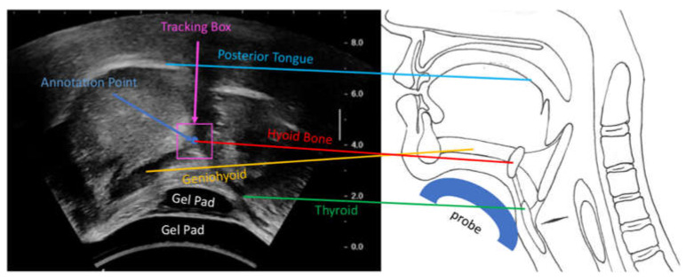
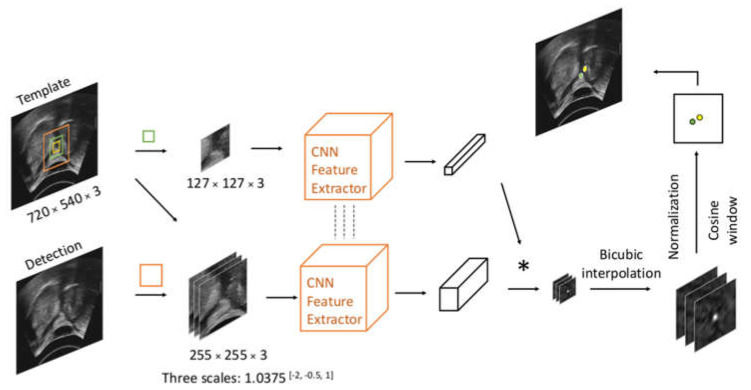
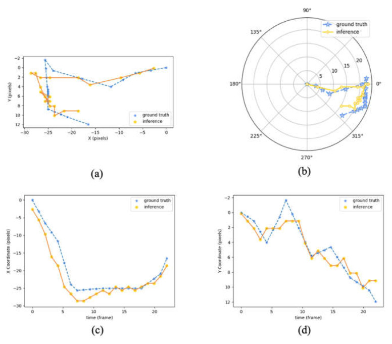
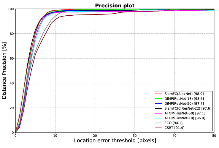
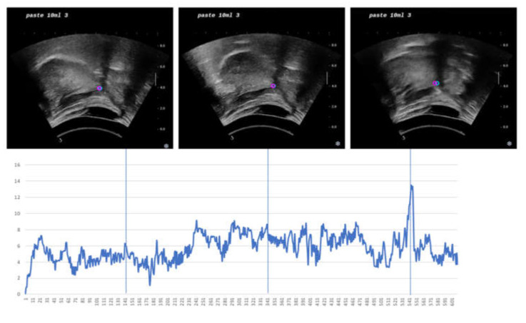
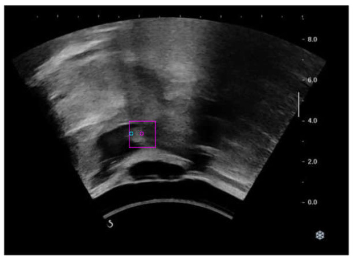
(1) Background: Ultrasound provides a radiation-free and portable method for assessing swallowing. Hyoid bone locations and displacements are often used as important indicators for the evaluation of swallowing disorders. However, this requires clinicians to spend a great deal of time reviewing the ultrasound images. (2) Methods: In this study, we applied tracking algorithms based on deep learning and correlation filters to detect hyoid locations in ultrasound videos collected during swallowing. Fifty videos were collected from 10 young, healthy subjects for training, evaluation, and testing of the trackers. (3) Results: The best performing deep learning algorithm, Fully-Convolutional Siamese Networks (SiamFC), proved to have reliable performance in getting accurate hyoid bone locations from each frame of the swallowing ultrasound videos. While having a real-time frame rate (175 fps) when running on an RTX 2060, SiamFC also achieved a precision of 98.9% at the threshold of 10 pixels (3.25 mm) and 80.5% at the threshold of 5 pixels (1.63 mm). The tracker's root-mean-square error and average error were 3.9 pixels (1.27 mm) and 3.3 pixels (1.07 mm), respectively. (4) Conclusions: Our results pave the way for real-time automatic tracking of the hyoid bone in ultrasound videos for swallowing assessment.
Keywords: SiamFC; correlation filters; deep learning; dysphagia; hyoid bone; real-time; swallowing; tracking; ultrasound videos.
Conflict of interest statement
The authors declare no conflict of interest. The funders had no role in the design of the study; in the collection, analyses, or interpretation of data; in the writing of the manuscript, or in the decision to publish the results.
Figures
Similar articles
-
Methodological Procedures to Acquire and Analyze Ultrasound Images of Swallowing: A Scoping Review.Dysphagia. 2025 Feb;40(1):1-53. doi: 10.1007/s00455-024-10714-1. Epub 2024 May 27. Dysphagia. 2025. PMID: 38802587
-
Deep Learning for Automatic Hyoid Tracking in Videofluoroscopic Swallow Studies.Dysphagia. 2023 Feb;38(1):171-180. doi: 10.1007/s00455-022-10438-0. Epub 2022 Apr 28. Dysphagia. 2023. PMID: 35482213
-
Automated assessment of hyoid movement during normal swallow using ultrasound.Int J Lang Commun Disord. 2022 May;57(3):615-629. doi: 10.1111/1460-6984.12712. Epub 2022 Mar 14. Int J Lang Commun Disord. 2022. PMID: 35285113 Free PMC article.
-
Automatic Tracking of Hyoid Bone Displacement and Rotation Relative to Cervical Vertebrae in Videofluoroscopic Swallow Studies Using Deep Learning.J Imaging Inform Med. 2024 Aug;37(4):1922-1932. doi: 10.1007/s10278-024-01039-4. Epub 2024 Feb 21. J Imaging Inform Med. 2024. PMID: 38383805 Free PMC article.
-
Quantitative approach to analyze hyoid bone movement during swallowing by ultrasound: an integrative review.Codas. 2023 Jul 17;35(4):e20220002. doi: 10.1590/2317-1782/20232022002pt. eCollection 2023. Codas. 2023. PMID: 37466503 Free PMC article. Review.
Cited by
-
AI-Based Detection of Aspiration for Video-Endoscopy with Visual Aids in Meaningful Frames to Interpret the Model Outcome.Sensors (Basel). 2022 Dec 4;22(23):9468. doi: 10.3390/s22239468. Sensors (Basel). 2022. PMID: 36502169 Free PMC article.
-
Translating Ultrasound into Clinical Practice for the Assessment of Swallowing and Laryngeal Function: A Speech and Language Pathology-Led Consensus Study.Dysphagia. 2022 Dec;37(6):1586-1598. doi: 10.1007/s00455-022-10413-9. Epub 2022 Feb 24. Dysphagia. 2022. PMID: 35201387 Free PMC article.
-
Hyoid Bone Movement During Swallowing in Female Thyroidectomy Patients: A Kinematic Ultrasound Study.Dysphagia. 2024 Oct;39(5):956-963. doi: 10.1007/s00455-024-10676-4. Epub 2024 Mar 4. Dysphagia. 2024. PMID: 38436670
-
Methodological Procedures to Acquire and Analyze Ultrasound Images of Swallowing: A Scoping Review.Dysphagia. 2025 Feb;40(1):1-53. doi: 10.1007/s00455-024-10714-1. Epub 2024 May 27. Dysphagia. 2025. PMID: 38802587
-
Visualizing tactile feedback: an overview of current technologies with a focus on ultrasound elastography.Front Med Technol. 2023 Oct 3;5:1238129. doi: 10.3389/fmedt.2023.1238129. eCollection 2023. Front Med Technol. 2023. PMID: 37854637 Free PMC article. Review.
References
-
- Smithard D.G. Dysphagia: A Geriatric Giant? Med. Clin. Rev. 2016;2:1–7. doi: 10.21767/2471-299X.1000014. - DOI
MeSH terms
Grants and funding
LinkOut - more resources
Full Text Sources
Medical
Miscellaneous
