miR-21-5p Regulates the Proliferation and Differentiation of Skeletal Muscle Satellite Cells by Targeting KLF3 in Chicken
- PMID: 34073601
- PMCID: PMC8227323
- DOI: 10.3390/genes12060814
miR-21-5p Regulates the Proliferation and Differentiation of Skeletal Muscle Satellite Cells by Targeting KLF3 in Chicken
Abstract
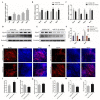
The proliferation and differentiation of skeletal muscle satellite cells (SMSCs) play an important role in the development of skeletal muscle. Our previous sequencing data showed that miR-21-5p is one of the most abundant miRNAs in chicken skeletal muscle. Therefore, in this study, the spatiotemporal expression of miR-21-5p and its effects on skeletal muscle development of chickens were explored using in vitro cultured SMSCs as a model. The results in this study showed that miR-21-5p was highly expressed in the skeletal muscle of chickens. The overexpression of miR-21-5p promoted the proliferation of SMSCs as evidenced by increased cell viability, increased cell number in the proliferative phase, and increased mRNA and protein expression of proliferation markers including PCNA, CDK2, and CCND1. Moreover, it was revealed that miR-21-5p promotes the formation of myotubes by modulating the expression of myogenic markers including MyoG, MyoD, and MyHC, whereas knockdown of miR-21-5p showed the opposite result. Gene prediction and dual fluorescence analysis confirmed that KLF3 was one of the direct target genes of miR-21-5p. We confirmed that, contrary to the function of miR-21-5p, KLF3 plays a negative role in the proliferation and differentiation of SMSCs. Si-KLF3 promotes cell number and proliferation activity, as well as the cell differentiation processes. Our results demonstrated that miR-21-5p promotes the proliferation and differentiation of SMSCs by targeting KLF3. Collectively, the results obtained in this study laid a foundation for exploring the mechanism through which miR-21-5p regulates SMSCs.
Keywords: miR-21-5p; muscle development; myogenic markers; myotubes.
Conflict of interest statement
The authors declare no conflict of interest.
Figures


Similar articles
-
miR-99a-5p Regulates the Proliferation and Differentiation of Skeletal Muscle Satellite Cells by Targeting MTMR3 in Chicken.Genes (Basel). 2020 Mar 29;11(4):369. doi: 10.3390/genes11040369. Genes (Basel). 2020. PMID: 32235323 Free PMC article.
-
miR-194-5p negatively regulates the proliferation and differentiation of rabbit skeletal muscle satellite cells.Mol Cell Biochem. 2021 Jan;476(1):425-433. doi: 10.1007/s11010-020-03918-0. Epub 2020 Sep 30. Mol Cell Biochem. 2021. PMID: 32997306 Free PMC article.
-
miR-9-5p Inhibits Skeletal Muscle Satellite Cell Proliferation and Differentiation by Targeting IGF2BP3 through the IGF2-PI3K/Akt Signaling Pathway.Int J Mol Sci. 2020 Feb 28;21(5):1655. doi: 10.3390/ijms21051655. Int J Mol Sci. 2020. PMID: 32121275 Free PMC article.
-
MicroRNA-381 Regulates Proliferation and Differentiation of Caprine Skeletal Muscle Satellite Cells by Targeting PTEN and JAG2.Int J Mol Sci. 2022 Nov 5;23(21):13587. doi: 10.3390/ijms232113587. Int J Mol Sci. 2022. PMID: 36362373 Free PMC article.
-
The roles of miRNAs in adult skeletal muscle satellite cells.Free Radic Biol Med. 2023 Nov 20;209(Pt 2):228-238. doi: 10.1016/j.freeradbiomed.2023.10.403. Epub 2023 Oct 24. Free Radic Biol Med. 2023. PMID: 37879420 Free PMC article. Review.
Cited by
-
Identification of crucial circRNAs in skeletal muscle during chicken embryonic development.BMC Genomics. 2022 Apr 28;23(1):330. doi: 10.1186/s12864-022-08588-4. BMC Genomics. 2022. PMID: 35484498 Free PMC article.
-
Transcriptome-Based Identification of the Muscle Tissue-Specific Expression Gene CKM and Its Regulation of Proliferation, Apoptosis and Differentiation in Chicken Primary Myoblasts.Animals (Basel). 2023 Jul 14;13(14):2316. doi: 10.3390/ani13142316. Animals (Basel). 2023. PMID: 37508090 Free PMC article.
-
Strand-Specific RNA Sequencing Reveals Gene Expression Patterns in F1 Chick Breast Muscle and Liver after Hatching.Animals (Basel). 2024 Apr 29;14(9):1335. doi: 10.3390/ani14091335. Animals (Basel). 2024. PMID: 38731340 Free PMC article.
-
Genome-Wide Analysis of the KLF Gene Family in Chicken: Characterization and Expression Profile.Animals (Basel). 2023 Apr 22;13(9):1429. doi: 10.3390/ani13091429. Animals (Basel). 2023. PMID: 37174466 Free PMC article.
-
Exploration of Potential Target Genes of miR-24-3p in Chicken Myoblasts by Transcriptome Sequencing Analysis.Genes (Basel). 2023 Sep 5;14(9):1764. doi: 10.3390/genes14091764. Genes (Basel). 2023. PMID: 37761904 Free PMC article.
References
-
- Chang N.C., Rudnicki M.A. Satellite cells: The architects of skeletal muscle. Curr. Top. Dev. Biol. 2014;107:161–181. - PubMed
Publication types
MeSH terms
Substances
LinkOut - more resources
Full Text Sources
Research Materials
Miscellaneous

