. 2021 May 26;9(6):609.
doi: 10.3390/biomedicines9060609.

Occlusion of the Superior Mesenteric Artery in Rats Reversed by Collateral Pathways Activation: Gastric Pentadecapeptide BPC 157 Therapy Counteracts Multiple Organ Dysfunction Syndrome; Intracranial, Portal, and Caval Hypertension; and Aortal Hypotension

Affiliations

Occlusion of the Superior Mesenteric Artery in Rats Reversed by Collateral Pathways Activation: Gastric Pentadecapeptide BPC 157 Therapy Counteracts Multiple Organ Dysfunction Syndrome; Intracranial, Portal, and Caval Hypertension; and Aortal Hypotension

Mario Knezevic et al. Biomedicines. .

Abstract

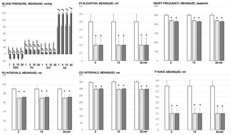
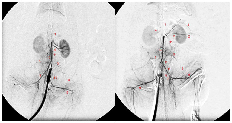
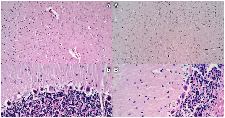
Gastric pentadecapeptide BPC 157 therapy counteracts multiple organ dysfunction syndrome in rats, which have permanent occlusion of the superior mesenteric artery close to the abdominal aorta. Previously, when confronted with major vessel occlusion, its effect would rapidly activate collateral vessel pathways and resolve major venous occlusion syndromes (Pringle maneuver ischemia, reperfusion, Budd-Chiari syndrome) in rats. This would overwhelm superior mesenteric artery permanent occlusion, and result in local, peripheral, and central disturbances. Methods: Assessments, for 30 min (gross recording, angiography, ECG, pressure, microscopy, biochemistry, and oxidative stress), included the portal hypertension, caval hypertension, and aortal hypotension, and centrally, the superior sagittal sinus hypertension; systemic arterial and venous thrombosis; ECG disturbances; MDA-tissue increase; and multiple organ lesions and disturbances, including the heart, lung, liver, kidney, and gastrointestinal tract, in particular, as well as brain (cortex (cerebral, cerebellar), hypothalamus/thalamus, hippocampus). BPC 157 therapy (/kg, abdominal bath) (10 µg, 10 ng) was given for a 1-min ligation time. Results: BPC 157 rapidly recruits collateral vessels (inferior anterior pancreaticoduodenal artery and inferior mesenteric artery) that circumvent occlusion and ascertains blood flow distant from the occlusion in the superior mesenteric artery. Portal and caval hypertension, aortal hypotension, and, centrally, superior sagittal sinus hypertension were attenuated or eliminated, and ECG disturbances markedly mitigated. BPC 157 therapy almost annihilated venous and arterial thrombosis. Multiple organ lesions and disturbances (i.e., heart, lung, liver, and gastrointestinal tract, in particular, as well as brain) were largely attenuated. Conclusions: Rats with superior mesenteric artery occlusion may additionally undergo BPC 157 therapy as full counteraction of vascular occlusion-induced multiple organ dysfunction syndrome.

Keywords: BPC 157; rats; superior mesenteric artery occlusion; vascular recruitment.


Conflict of interest statement

The authors declare that there are no conflict of interest.

Figures

Figure 1
Figure 1
Animal preparation for the assessment procedure. (a). For superior sagittal sinus pressure recording, a single burr hole was placed in the rostral part of the sagittal suture, above the superior sagittal sinus (arrow); (b). Six burr holes drilled in three horizontal lines, with all of them medial to the superior temporal lines and temporalis muscle attachments; (c). Complete calvariectomy.
Figure 2
Figure 2
Mucosal lesions in the stomach, duodenum, jejunum, cecum, and ascending colon (sum of the longest lesions’ diameters, mm, means ± SD, mm, at 30 min of ligation), and the serosal disturbance in the stomach, duodenum, jejunum, cecum, and ascending colon, as hemorrhage, congestion, arterial filling, and ramification, scored 0–4, min/med/max) at 1, 5, 15, and 30 min after medication application (10 µg/kg BPC 157 (light gray bars), 10 ng/kg BPC 157 (dark gray bars), or 5 mL/kg saline (white bars). * p ˂ 0.05, at least, vs. control.
Figure 3
Figure 3
Proportional change of the vessel surface area assessed for failure of peripheral vessels’ development, as % of volume in healthy, means ± SD, mm), abdominal aorta, inferior mesenteric artery, inferior caval vein, and superior mesenteric artery relative volume at the time of the occlusion, proximal to ligation and distal to ligation, before medication application (SMA occlusion before medication) (white bars) and at 1, 5, 15, and 30 min after medication application (10 µg/kg BPC 157 (light gray bars), 10 ng/kg BPC 157 (dark gray bars), or 5mL/kg saline (white bars) (SMA occlusion after medication). All values significantly different (p ˂ 0.05, at least) vs. healthy values (100%); * p ˂ 0.05, at least, vs. corresponding control.
Figure 4
Figure 4
Rats with an occluded superior mesenteric artery and that received saline (small letters, ae) or BPC 157 therapy (capitals, AE) showed quite distinctive presentation of the brain at the end of the experiments (a,A), as well as the superior mesenteric artery (b,B), inferior caval vein and aorta (c,C), inferior anterior pancreaticoduodenal artery (d,D), and inferior mesenteric artery and its ramification (e,E) soon after mediation application. Arrows indicate important points. Arrows show ligation (b,B), and the distal part of the superior mesenteric artery, filled with blood (B) or thin and empty in controls (b), in addition to the superior mesenteric vein, congested (controls, b) and non-congested (BPC 157-rats, B). Arrows show the congested inferior caval vein and thin abdominal aorta (controls, c) and non-congested inferior caval vein and abdominal aorta with a maintained volume (BPC 157-rats, C). Arrows show the congested inferior anterior pancreaticoduodenal vein with no concurrent presentation of the inferior anterior pancreaticoduodenal artery along with the vein, and an apparent gape, and no communication with the superior anterior pancreaticoduodenal artery (controls, d), in contrast to the non-congested inferior anterior pancreaticoduodenal vein with clear presentation of the inferior anterior pancreaticoduodenal artery along with the vein, and clear communication with the superior anterior pancreaticoduodenal artery (BPC 157-treated rats, D). Arrows show the empty and thin inferior mesenteric artery (controls, e), and the inferior mesenteric artery filled with blood and its ramification (BPC 157-rats, E). The camera was attached to a VMS-004 Discovery Deluxe USB microscope (Veho, Denver, CO, USA).
Figure 5
Figure 5
Rats with an occluded superior mesenteric artery and that received saline (small letters, ae) or BPC 157 therapy (capitals, AE) showed quite a distinctive mucosal presentation of the stomach (a,A), duodenum (b,B), jejunum (c,C), cecum (d,D), and ascending colon (e,E) at the end of the experiments, with evident lesions in controls (low) and preserved mucosa integrity in BPC 157-treated rats (upper). The camera was attached to a VMS-004 Discovery Deluxe USB microscope (Veho, Denver, CO, USA).
Figure 6
Figure 6
Timeline of the brain presentation in rats after complete calvariectomy: normal brain before superior mesenteric artery ligation (healthy, h), brain swelling immediately after occlusion of the superior mesenteric artery (a), and after BPC 157 therapy, a gradual decrease of the swelling, immediately after therapy (b), and at 5 min (c) and 15 min (d) thereafter. The camera was attached to a VMS-004 Discovery Deluxe USB microscope (Veho, Denver, CO, USA). Proportional change of the brain surface area assessed for the brain-swelling recording, as % of volume in healthy means ± SD, mm), brain, abdominal aorta, inferior mesenteric artery, inferior caval vein, and superior mesenteric artery relative to the volume at the time of the occlusion (SMA occlusion before medication) (white bars) and at 1, 5, 15, and 30 min after medication application (10 µg/kg BPC 157 (light gray bars), 10 ng/kg BPC 157 (dark gray bars), or 5 mL/kg saline (white bars) (SMA occlusion after medication). # p ˂ 0.05, at least, vs. healthy values (100%); * p ˂ 0.05, at least, vs. corresponding control.
Figure 7
Figure 7
Blood pressure (mmHg) in the superior sagittal sinus (SSS), portal vein (PV), inferior caval vein (ICV), and abdominal aorta (AA), and ECG disturbance, ST elevation (mV), heart frequency (beats/min), PQ intervals (ms), QTc intervals (ms), and p wave amplitude (mV), mm, means ± SD) at 1, 5, 15, and 30 min after medication application (10 µg/kg BPC 157 (light gray bars), 10 ng/kg BPC 157 (dark gray bars), or 5 mL/kg saline (white bars). * p ˂ 0.05, at least, vs. control.
Figure 8
Figure 8
Gastrointestinal lesions (scored 0–15), scoring of the lesions in the lung (0–18), liver (0–9), kidney (0–12), heart (0–3), and brain (cerebral cortex, cerebellar cortex min/med/max thrombus mass (g)) in the superior sagittal sinus (SSS), portal vein (PV), superior mesenteric artery (SMA), inferior caval vein (ICV), superior mesenteric vein (SMV), hepatic artery (HA), and abdominal aorta (AA). Blood pressure (mmHg) in the superior sagittal sinus (SSS), portal vein (PV), inferior caval vein (ICV), and abdominal aorta (AA), means ± SD, serum enzymes (ALT, AST) values (U/L), means ± SD, and MDA values (µmol/mg protein) in cecum, means ± SD, at the end of the experiment in the rats with occlusion of the superior mesenteric artery and that received medication application (10 µg/kg BPC 157 (light gray bars), 10 ng/kg BPC 157 (dark gray bars), or 5mL/kg saline (white bars). * p ˂ 0.05, at least, vs. control.
Figure 9
Figure 9
Angiography was performed in rats with ligation of the superior mesenteric artery at 15 min post-ligation, using a C-VISION PLUS fluoroscopy unit (Shimadzu, Japan). One milliliter, for 45 s, of warmed Omnipaque 350 (iohexol) non-ionic contrast medium (GE Healthcare, USA) was injected into the abdominal aorta at the level of the lumbar arteries. Presentation of the abdominal aorta (0), coeliac trunk (1), superior mesenteric artery (2), inferior anterior pancreaticoduodenal artery (3), renal artery (4), iliolumbar artery (5), common iliac artery (6), internal iliac artery (7), caudal epigastric artery (8), iliacofemoral artery (9), testicular artery (10), and inferior mesenteric artery (11) in rats (Low et al., 2016) with occlusion of the superior mesenteric artery, immediately after they received medication application (10 µg/kg BPC 157 (right) or 5 mL/kg saline (left) at 15 min of ligation.
Figure 10
Figure 10
Stomach and duodenal injury (HE x100). Control rats show marked transmural congestion within the lamina propria of the stomach (a, left, upper) and duodenum (b, left, low). Blunt duodenal villi and mild hyperplasia of the crypts. Contrarily, BPC 157 rats exhibit only mild congestion, with preserved stomach (A, right, upper) and duodenal (B, right, low) architecture of the mucosa.
Figure 11
Figure 11
Intestinal injury (HE, ×100 (upper), ×200 (low)). Controls presented with moderately severe mucosal injury with a reduction of intestinal villi; more severe mucosal injury with lumen dilatation of the colon and a reduction of crypts (a,b, left). Contrarily, BPC 157-treated rats exhibited only mild congestion, with preserved mucosal intestinal architecture (A,B, right).
Figure 12
Figure 12
Congestion and hemorrhage of the heart and lung. (HE, ×100). Control rats exhibit congestion within the myocardium (a, left, upper) and lung septa along with intralveolar hemorrhage; lung edema and focal accumulation of neutrophils within septa (b, left, low). Contrarily, no heart congestion (A, right, upper) and no lung congestion and no intralveolar hemorrhage (B, right, low) were observed in BPC 157-treated rats.
Figure 13
Figure 13
Liver and kidney injury (HE, ×100). In liver parenchyma, control rats showed congestion and dilatation of the sinusoids and central veins (a, left, upper) while BPC 157 rats showed no changes in liver parenchyma (A, right, upper). Controls showed congestion of the renal parenchyma along with congestion of the glomeruli (b, left, low); BPC 157 rats showed no changes in renal parenchyma (B, right, low).
Figure 14
Figure 14
Brain histology. Control rats presented increased edema and congestion and an increased number of karyopyknotic cells in the cerebral (a) (HE, ×200) and cerebellar cortex (b) (HE, ×400). Contrarily, BPC 157-treated rats exhibited a few karyopyknotic neuronal cells in the cerebral (A) (HE, ×200) and cerebellar cortex (B) (HE, ×400).
Figure 15
Figure 15
Brain histology: Control rats presented increased edema and congestion and an increased number of karyopyknotic cells in the hippocampus cortex (a) and hypothalamus (b), (HE, ×200). Contrarily, BPC 157-treated rats exhibited a few karyopyknotic neuronal cells in the hippocampus cortex (A) and hypothalamus (B) (HE, ×200).

Similar articles

Cited by

References

    1. Sikiric P., Petek M., Rucman R., Seiwerth S., Grabarevic Z., Rotkvic I., Turkovic B., Jagic V., Mildner B., Duvnjak M., et al. A new gastric juice peptide, BPC. An overview of the stomach-stress-organoprotection hypothesis and beneficial effects of BPC. J. Physiol. Paris. 1993;87:313–327. doi: 10.1016/0928-4257(93)90038-U. - DOI - PubMed
    1. Sikiric P., Seiwerth S., Brcic L., Blagaic A.B., Zoricic I., Sever M., Klicek R., Radic B., Keller N., Sipos K., et al. Stable gastric pentadecapeptide BPC 157 in trials for inflammatory bowel disease (PL-10, PLD-116, PL 14736, Pliva, Croatia). Full and distended stomach, and vascular response. Inflammopharmacology. 2006;14:214–221. doi: 10.1007/s10787-006-1531-7. - DOI - PubMed
    1. Sikiric P., Seiwerth S., Brcic L., Sever M., Klicek R., Radic B., Drmic D., Ilic S., Kolenc D. Revised Robert’s cytoprotection and adaptive cytoprotection and stable gastric pentadecapeptide BPC 157. Possible significance and implications for novel mediator. Curr. Pharm. Des. 2010;16:1224–1234. doi: 10.2174/138161210790945977. - DOI - PubMed
    1. Sikiric P., Seiwerth S., Rucman R., Turkovic B., Rokotov D.S., Brcic L., Sever M., Klicek R., Radic B., Drmic D., et al. Stable gastric pentadecapeptide BPC 157: Novel therapy in gastrointestinal tract. Curr. Pharm. Des. 2011;17:1612–1632. doi: 10.2174/138161211796196954. - DOI - PubMed
    1. Sikiric P., Seiwerth S., Rucman R., Turkovic B., Brcic L., Sever M., Klicek R., Radic B., Drmic D., Ilic S., et al. Focus on ulcerative colitis: Stable gastric pentadecapeptide BPC 157. Curr. Med. Chem. 2012;19:126–132. doi: 10.2174/092986712803414015. - DOI - PubMed

LinkOut - more resources