Review
. 2021 May 26;11(6):959.
doi: 10.3390/diagnostics11060959.

Artificial Intelligence Based Algorithms for Prostate Cancer Classification and Detection on Magnetic Resonance Imaging: A Narrative Review

Affiliations
Review

Artificial Intelligence Based Algorithms for Prostate Cancer Classification and Detection on Magnetic Resonance Imaging: A Narrative Review

Jasper J Twilt et al. Diagnostics (Basel). .

Abstract

Due to the upfront role of magnetic resonance imaging (MRI) for prostate cancer (PCa) diagnosis, a multitude of artificial intelligence (AI) applications have been suggested to aid in the diagnosis and detection of PCa. In this review, we provide an overview of the current field, including studies between 2018 and February 2021, describing AI algorithms for (1) lesion classification and (2) lesion detection for PCa. Our evaluation of 59 included studies showed that most research has been conducted for the task of PCa lesion classification (66%) followed by PCa lesion detection (34%). Studies showed large heterogeneity in cohort sizes, ranging between 18 to 499 patients (median = 162) combined with different approaches for performance validation. Furthermore, 85% of the studies reported on the stand-alone diagnostic accuracy, whereas 15% demonstrated the impact of AI on diagnostic thinking efficacy, indicating limited proof for the clinical utility of PCa AI applications. In order to introduce AI within the clinical workflow of PCa assessment, robustness and generalizability of AI applications need to be further validated utilizing external validation and clinical workflow experiments.

Keywords: artificial intelligence; computer-aided diagnosis; deep learning; machine learning; magnetic resonance imaging; prostate neoplasms; radiomics.


Conflict of interest statement

J.J.T., K.G.v.L. and M.d.R. declare no relationships with any companies or services related to the subject matter of this article. J.J.F. and H.J.H. receive research grants from Siemens Healthineers. The funders had no role in the design of the study; in the collection, analyses, or interpretation of data; in the writing of the manuscript, or in the decision to publish the results.

Figures
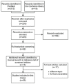
Figure 1
Figure 1
Flow diagram for search strategy.
Figure 2
Figure 2
Overview of number of studies and commercially available AI applications for prostate MRI included in this review between 2018 and February 2021. Studies are categorized according to two-class lesion classification with machine learning (ML) and deep learning (DL), multi-class lesion classification, two-class lesion detection, and multi-class lesion detection. Most studies were observed for ML based two-class lesion characterization.
Figure 3
Figure 3
Machine learning (ML) workflow of two-class lesion classification for prostate cancer using an axial T2-weighted sequence. As input, multiparametric or singular MR sequences are used. Regions of interests (ROIs) are annotated, labeled, and used for feature extraction. A selection of features is used to train the ML-algorithm. As output, the annotated region is classified in one of the two classes.
Figure 4
Figure 4
Deep learning (DL) workflow of two-class lesion classification for prostate cancer using an axial T2-weighted sequence. As input, multiparametric or singular MR sequences are used. On the MR images, regions of interest (ROIs) (patches or volumes) are annotated. The patches and/or ROIs are fed into the DL-algorithm. As output, a predicted label for the corresponding patch and/or ROI is provided.
Figure 5
Figure 5
Workflow of machine learning (ML) and deep learning (DL) based multi-class lesion classification for prostate cancer using an axial T2-weighted sequence. The workflow follows a similar workflow as ML and DL pipelines described within two-class classification (see Figure 3 and Figure 4). As input, multiparametric or single MR sequences are utilized. Regions of interest (ROIs) are annotated and feature selection may be implemented prior to algorithm training. Classification is divided into multiple classes utilizing multiple labels within the ML and DL algorithm output. As output, annotations are graded according to the various labels (groups 1, 2, 3… n).
Figure 6
Figure 6
Deep learning (DL) and machine learning (ML) workflow of algorithms for two-class lesion detection for prostate cancer (PCa) using an axial T2-weighted sequence. As input, multiparametric or single MR sequences are utilized. During this, training features are trained and used to classify image voxels within benign or malignant classes. Algorithms provide a probability map for prostate cancer likelihood. Based on a threshold within the probability map (e.g., probability > 0.5), prostate cancer segmentations (red) or attention boxes based on prostate cancer segmentations (yellow) may be extracted.

References

    1. Sung H., Ferlay J., Siegel R.L., Laversanne M., Soerjomataram I., Jemal A., Bray F. Global cancer statistics 2020: GLOBOCAN estimates of incidence and mortality worldwide for 36 cancers in 185 countries. CA Cancer J. Clin. 2021;68:394–424. doi: 10.3322/caac.21660. - DOI - PubMed
    1. Siegel R.L., Miller K.D., Jemal A. Cancer statistics, 2020. CA Cancer J. Clin. 2020;70:7–30. doi: 10.3322/caac.21590. - DOI - PubMed
    1. Martín Noguerol T., Paulano-Godino F., Martín-Valdivia M.T., Menias C.O., Luna A. Strengths, Weaknesses, Opportunities, and Threats Analysis of Artificial Intelligence and Machine Learning Applications in Radiology. J. Am. Coll. Radiol. 2019;16:1239–1247. doi: 10.1016/j.jacr.2019.05.047. - DOI - PubMed
    1. Kasivisvanathan V., Rannikko A.S., Borghi M. MRI-targeted or standard biopsy for prostate-cancer diagnosis. N. Engl. J. Med. 2018;378:1767–1777. doi: 10.1056/NEJMoa1801993. - DOI - PMC - PubMed
    1. Rouvière O., Puech P., Renard-Penna R., Claudon M., Roy C., Mège-Lechevallier F., Decaussin-Petrucci M., Dubreuil-Chambardel M., Magaud L., Remontet L., et al. Use of prostate systematic and targeted biopsy on the basis of multiparametric MRI in biopsy-naive patients (MRI-FIRST): A prospective, multicentre, paired diagnostic study. Lancet Oncol. 2019;20:100–109. doi: 10.1016/S1470-2045(18)30569-2. - DOI - PubMed

Grants and funding

LinkOut - more resources