. 2021 May 24;22(11):5511.
doi: 10.3390/ijms22115511.

Sex-Specific Cannabidiol- and Iloperidone-Induced Neuronal Activity Changes in an In Vitro MAM Model System of Schizophrenia

Affiliations

Sex-Specific Cannabidiol- and Iloperidone-Induced Neuronal Activity Changes in an In Vitro MAM Model System of Schizophrenia

Rachel-Karson Thériault et al. Int J Mol Sci. .

Abstract

Cortical circuit dysfunction is thought to be an underlying mechanism of schizophrenia (SZ) pathophysiology with normalization of aberrant circuit activity proposed as a biomarker for antipsychotic efficacy. Cannabidiol (CBD) shows potential as an adjunctive antipsychotic therapy; however, potential sex effects in these drug interactions remain unknown. In the present study, we sought to elucidate sex effects of CBD coadministration with the atypical antipsychotic iloperidone (ILO) on the activity of primary cortical neuron cultures derived from the rat methylazoxymethanol acetate (MAM) model used for the study of SZ. Spontaneous network activity measurements were obtained using a multielectrode array at baseline and following administration of CBD or ILO alone, or combined. At baseline, MAM male neurons displayed increased bursting activity whereas MAM female neurons exhibited no difference in bursting activity compared to sex-matched controls. CBD administered alone showed a rapid but transient increase in neuronal activity in the MAM networks, an effect more pronounced in females. Furthermore, ILO had an additive effect on CBD-induced elevations in activity in the MAM male neurons. In the MAM female neurons, CBD or ILO administration resulted in time-dependent elevations in neuronal activity, but the short-term CBD-induced increases in activity were lost when CBD and ILO were combined. Our findings indicate that CBD induces rapid increases in cortical neuronal activity, with sex-specific drug interactions upon ILO coadministration. This suggests that sex should be a consideration when implementing adjunct therapy for treatment of SZ.

Keywords: cannabidiol; electrophysiology; haloperidol; iloperidone; primary cortical neurons; schizophrenia.


Conflict of interest statement

The authors declare no conflict of interest.

Figures
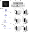
Figure 1
Figure 1
Effect of MAM on neuronal cortical systems activity. (A) Representative immunostaining of MAP2 showing a DIV 21 primary cortical neuron. (B) Western blot showing levels of Syn1, PSD-95 and actin at DIV 7, 14, 21, and 28 in primary cortical neurons derived from male and female SAL treated (control) and MAM rats. (C) Representative raster plots showing sex differences in baseline neuronal activity. (D) Quantification of group differences in neuronal firing rate showed that female SAL and MAM rat neurons exhibited reduced firing rates at baseline compared to male neurons of the same model. (E,F) Male MAM-derived neurons exhibited increased number of bursts compared to male SAL neurons, whereas female MAM neurons exhibited no difference in bursting activity compared to female SAL neurons. (G) Inter-burst interval showing a shorter interval in MAM-derived neurons. (H) Female SAL neurons showed a decreased synchrony index at baseline. (I) Male MAM-derived neurons exhibited elevated network bursting compared to all other groups. Data are expressed as means ± SEM. N = 3 biological replicates. * p < 0.05, ** p < 0.01, compared to males of the same model, # p < 0.5, ## p < 0.01, ### p < 0.001 compared to sex-matched controls, ANOVA followed by Bonferroni or Games–Howell post-hoc determined by Levene’s test of variance.
Figure 2
Figure 2
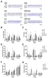
Temporal effect of cannabidiol (CBD) and/or antipsychotic exposure on neuronal activity in cultures derived from male SAL rats. (A) Representative raster plots showing neuronal activity following administration of each drug at 5 min, 20 min, and 24 h. (B) CBD or CBD+iloperidone (ILO) elevated spiking activity at 5 min, with a reduction in spiking by CBD at 24 h. (C) CBD alone or combined with haloperidol (HAL) or ILO increased the number of neuronal bursts at 5 min, an effect maintained in the CBD+HAL group at 20 min. At 24 h, CBD or CBD+ILO groups showed reduced number of bursts. (D) HAL administration reduced burst frequency at 5 min, whereas ILO increased bursting frequency at 20 min. At 24 h, CBD or CBD+ILO administration suppressed bursting frequency. (E) CBD alone, or with ILO, increased the inter-burst interval at 24 h. (F,G) Only CBD+ILO increased the synchrony index in male SAL neurons with no drug effects on the number of network bursts. Data are expressed as means ± SEM. N = 3 biological replicates. * p < 0.05, ** p < 0.01 compared to VEH-exposed neurons, planned comparison Student’s t-test.
Figure 3
Figure 3
Temporal effect of cannabidiol (CBD) and/or antipsychotic exposure on neuronal activity in cultures derived from female SAL rats. (A) Representative raster plots following administration of each drug at 5 min, 20 min, and 24 h. (BD) All drugs induced a transient decrease in firing rate, number of bursts, and burst frequency with all drugs inducing significantly reduced activity at 24 h. However, haloperidol (HAL) showed rapid onset effects, decreasing firing and bursting activity at 5 min. (EG) There were no drug effects on inter-burst interval, synchrony index, or the number of network bursts at any time point. Data are expressed as means ± SEM. N = 3 biological replicates. * p < 0.05, ** p < 0.01, *** p < 0.001 compared to VEH-exposed neurons, planned comparison Student’s t-test.
Figure 4
Figure 4
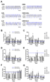
Temporal effect of cannabidiol (CBD) and/or antipsychotic exposure on neuronal activity in cultures derived from male MAM rats. (A) Representative raster plots following administration of each drug at 5 min, 20 min, and 24 h. (B) Compared to VEH-exposed neurons, at 5 min CBD increased the spiking rate, an effect exacerbated in the presence of iloperidone (ILO). At 24 h, CBD+haloperidol (HAL) administration lowered the spiking rate. (C) CBD+ILO increased the number of bursts at 5 min, with CBD alone suppressing burst number at 24 h. (D) CBD or CBD+ILO increased the bursting frequency at 5 min, with CBD or CBD+HAL lowering it at 24 h. (E) HAL alone resulted in a lower inter-burst interval at 5 min, whereas CBD+HAL elevated it at 20 min. (F) CBD+HAL suppressed the synchrony index at 5 min. (G) There were no drug effects on the number of network bursts. SAL- and MAM-derived baseline data is depicted by the blue and black dashed lines, respectively. Data are expressed as means ± SEM. N = 3 biological replicates. * p < 0.05, ** p < 0.01, *** p < 0.001 compared to VEH-exposed neurons, planned comparison Student’s t-test.
Figure 5
Figure 5
Temporal effect of cannabidiol (CBD) and/or antipsychotic exposure on neuronal activity in cultures derived from female MAM rats. (A) Representative raster plots following administration of each drug at 5 min, 20 min, and 24 h. (B) CBD exposure resulted in a higher firing rate at 5 min compared to VEH. Haloperidol (HAL) or iloperidone (ILO) increased the firing rate at 24 h. (C,D) HAL or CBD alone resulted in an elevated number of bursts and bursting frequency at 5 min. ILO administration also elevated the bursting frequency at this time point. At 24 h, HAL or ILO elevated both the number of bursts and the bursting frequency. (EG) There were no drug effects observed at any time point on the inter-burst interval, synchrony index, or number of network bursts. SAL- and MAM-derived baseline data is depicted by the blue and black dashed lines, respectively. Data are expressed as means ± SEM. N = 3 biological replicates. * p < 0.05, ** p < 0.01, *** p < 0.001 compared to VEH-exposed neurons, planned comparison Student’s t-test.

Similar articles

Cited by

References

    1. Marder S.R., Cannon T.D. Schizophrenia. N. Engl. J. Med. 2019;381:1753–1761. doi: 10.1056/NEJMra1808803. - DOI - PubMed
    1. American Psychiatric Association . Diagnostic and Statistical Manual of Mental Disorders. 5th ed. American Psychiatric Association; Washington, DC, USA: 2013. Schizophrenia and other psychotic disorders.
    1. Nielsen R.E., Levander S., Kjaersdam Telleus G., Jensen S.O., Ostergaard Christensen T., Leucht S. Second-generation antipsychotic effect on cognition in patients with schizophrenia—A meta-analysis of randomized clinical trials. Acta Psychiatr. Scand. 2015;131:185–196. doi: 10.1111/acps.12374. - DOI - PubMed
    1. Abel K.M., Drake R., Goldstein J.M. Sex differences in schizophrenia. Int. Rev. Psychiatry. 2010;22:417–428. doi: 10.3109/09540261.2010.515205. - DOI - PubMed
    1. Howard C.H., Fiedosewicz H., Patel C., Klegon D.A., Bayog R., Berman I. Treatment response and gender in patients with schizophrenia and schizoaffective disorder. Schizophr. Res. 2001;49:232.

MeSH terms