Review
. 2021 May 24;10(6):1297.
doi: 10.3390/cells10061297.

Co-Adaptation of Physical Attributes of the Mammalian Female Reproductive Tract and Sperm to Facilitate Fertilization

Affiliations
Review

Co-Adaptation of Physical Attributes of the Mammalian Female Reproductive Tract and Sperm to Facilitate Fertilization

Chih-Kuan Tung et al. Cells. .

Abstract

The functions of the female reproductive tract not only encompass sperm migration, storage, and fertilization, but also support the transport and development of the fertilized egg through to the birth of offspring. Further, because the tract is open to the external environment, it must also provide protection against invasive pathogens. In biophysics, sperm are considered "pusher microswimmers", because they are propelled by pushing fluid behind them. This type of swimming by motile microorganisms promotes the tendency to swim along walls and upstream in gentle fluid flows. Thus, the architecture of the walls of the female tract, and the gentle flows created by cilia, can guide sperm migration. The viscoelasticity of the fluids in the tract, such as mucus secretions, also promotes the cooperative swimming of sperm that can improve fertilization success; at the same time, the mucus can also impede the invasion of pathogens. This review is focused on how the mammalian female reproductive tract and sperm interact physically to facilitate the movement of sperm to the site of fertilization. Knowledge of female/sperm interactions can not only explain how the female tract can physically guide sperm to the fertilization site, but can also be applied for the improvement of in vitro fertilization devices.

Keywords: cervix; fertilization; oviduct; sperm; uterus; vagina.


Conflict of interest statement

The authors declare no conflict of interest. The funders had no role in the writing of the manuscript.

Figures

Figure 1
Figure 1
Basic anatomy of mammalian sperm, used with permission from Ref. [9]. Copyright 1975 Elsevier. In this drawing, the plasma membrane has been omitted in order to reveal internal structures. Cross sections are shown at various points along the length of the sperm, indicated by dashed lines. Mean lengths of sperm of mammalian species vary from 33.5 µm to 356.3 µm; the length of the head is less variable than the length of the flagellum, and averages roughly 8 µm [10].
Figure 2
Figure 2
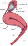
Left lateral view of the human female reproductive tract. The uterus, cervix and vagina have been bisected to show the entrance to the cervix and the uterotubal junction (UTJ). Only the right ovary and oviduct are shown. The ovarian ligament attaches the ovary to the uterus.
Figure 3
Figure 3
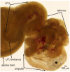
A transilluminated mouse oviduct. The ovary has been removed, but the coiling of the oviduct has been left in place. The size of mouse sperm (about 125 μm) is illustrated above the scale bar. To appreciate the size of the oocytes and cumulus in the ampulla, one of the oocytes has been covered by a solid black circle and the border of its cumulus indicated by a dotted line. Note that longitudinal folds line the uterotubal junction (UTJ) and the ampulla, while transversely oriented folds that form pockets can be seen in the isthmus, the site of sperm storage. Only the tip of a uterine horn is shown. Used with permission from Ref. [38]. Copyright 2010 Oxford University Press.
Figure 4
Figure 4
Tracks of sperm interactions with solid structures, as seen looking down from above. (a) Sperm that approach a sidewall from all angles (starting points denoted with open dots) swim along the sidewall (end points denoted with filled black dots) after hitting the wall. The location of wall contact is adjusted to (0,0) for each trajectory. (b) When sperm encounter a microgroove structure, the overwhelming majority of them remain in the groove. The starting points of the tracks are adjusted to (0,0). End points of the sperm trajectories are indicated by black dots. Note that sperm swim in either direction from the point of entry into the microgroove. Only one out of 50 sperm swam out of the microgroove. Adapted from Ref. [53]. Copyright 2015 by the authors.
Figure 5
Figure 5
Bull sperm response to fluid flow. The coordinate (0,0) marks the starting point of all of the tracks, which were followed for 2.8 s. (a) When there is no flow, sperm exhibit curved trajectories as a segment of their near-circular tracks. (b) When the flow rate is below the threshold for upstream swimming (which was measured as 1.1 µL/min), sperm trajectories drift downstream, i.e., the trajectories to the left are extended further and curved less than the ones to the right. (c) Once the flow rate is above the onset, sperm exhibit nearly linear trajectories with an upstream component. (a) and (c) adapted from Ref. [57]. Copyright 2015 American Physical Society. (b) adapted from Ref. [53]. Copyright 2015 by the authors.
Figure 6
Figure 6
Bull sperm collective swimming in viscoelastic fluid. (a) At lower sperm numbers and 1% PAM solution, sperm form nonbinding clusters (yellow ovals) in which several neighboring sperm swim in the same direction. (b) At higher sperm numbers and 0.7% PAM plus 1% PVP solution (PVP was added to increase the fluid viscosity to reduce thermal-like randomization), after a pulse of flow was applied and had dissipated, a mass of sperm swam toward the same direction.
Figure 7
Figure 7
Illustrations of sperm aggregation in three species. (a) A small sperm train, in which sperm from wood mice (Apodemus sylvaticus) were joined when the hooks on their heads latched onto flagella or hooks of neighboring sperm. (b) Rouleaux in sperm from guinea pigs (Cavia porcellus), in which heads are stacked together. (c) Shows how sperm from the grey short-tailed opossum (Monodelphis domestica) form pairs and how single sperm and pairs move in non-viscous and viscous media. Paired sperm swim progressively in non-viscous and viscous media, while single sperm only swim progressively in non-viscous medium (opossum sperm illustrations used with permission from Ref. [105]. Copyright 1995 Oxford University Press.).
Figure 8
Figure 8
A microfluidic device designed to model fluid flows and microgrooves within the cervix. (a) Diagrams of bull sperm and T. foetus. (b) Illustration of a bovine female reproductive tract (from Ref. [108]). UTJ (uterotubal junction). The pink arrow points in the direction of fluid flow through the cervix. (c) Microgrooves are seen in PAS/hematoxylin-stained frozen sections of bovine cervix. (d) Diagram of microfluidic device that recreates the microgrooves and fluid flows of the bovine cervix. The sperm seeding port is on the left side and the flow inlet on the right; they are connected by channels with and without microgrooves. Detail of the channel design in the middle of the device: (e) G denotes channels with microgrooves in the upper surface and F denotes a control channel without grooves. (f) A 3D drawing illustrates the details of grooved channels. The cross-sectional dimensions of the microgrooves are 20 μm × 20 μm (drawing not to scale). Adapted with permission from Ref. [36]. Copyright 2015 Springer Nature.

References

    1. Suarez S.S., Pacey A.A. Sperm transport in the female reproductive tract. Hum. Reprod. Update. 2006;12:23–37. doi: 10.1093/humupd/dmi047. - DOI - PubMed
    1. Yániz J.L., Lopez-Gatius F., Santolaria P., Mullins K.J. Study of the functional anatomy of bovine oviductal mucosa. Anat. Rec. 2000;260:268–278. doi: 10.1002/1097-0185(20001101)260:3<268::AID-AR60>3.0.CO;2-L. - DOI - PubMed
    1. Kölle S. Transport, Distribution and Elimination of Mammalian Sperm Following Natural Mating and Insemination. Reprod. Domest. Anim. 2015;50:2–6. doi: 10.1111/rda.12576. - DOI - PubMed
    1. Suarez S.S., Brockman K., Lefebvre R. Distribution of mucus and sperm in bovine oviducts after artificial insemination: The physical environment of the oviductal sperm reservoir. Biol. Reprod. 1997;56:447–453. doi: 10.1095/biolreprod56.2.447. - DOI - PubMed
    1. Mullins K.J., Saacke R.G. Study of the functional anatomy of bovine cervical mucosa with special reference to mucus secretion and sperm transport. Anat. Rec. 1989;225:106–117. doi: 10.1002/ar.1092250205. - DOI - PubMed

Publication types

LinkOut - more resources