. 2021 May 24;22(11):5523.
doi: 10.3390/ijms22115523.

Simulating Peptide Monolayer Formation: GnRH-I on Silica

Affiliations

Simulating Peptide Monolayer Formation: GnRH-I on Silica

Neret Pujol-Navarro et al. Int J Mol Sci. .

Abstract

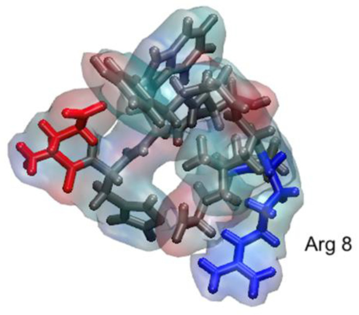
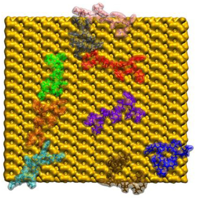
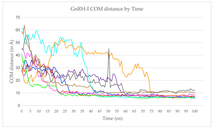
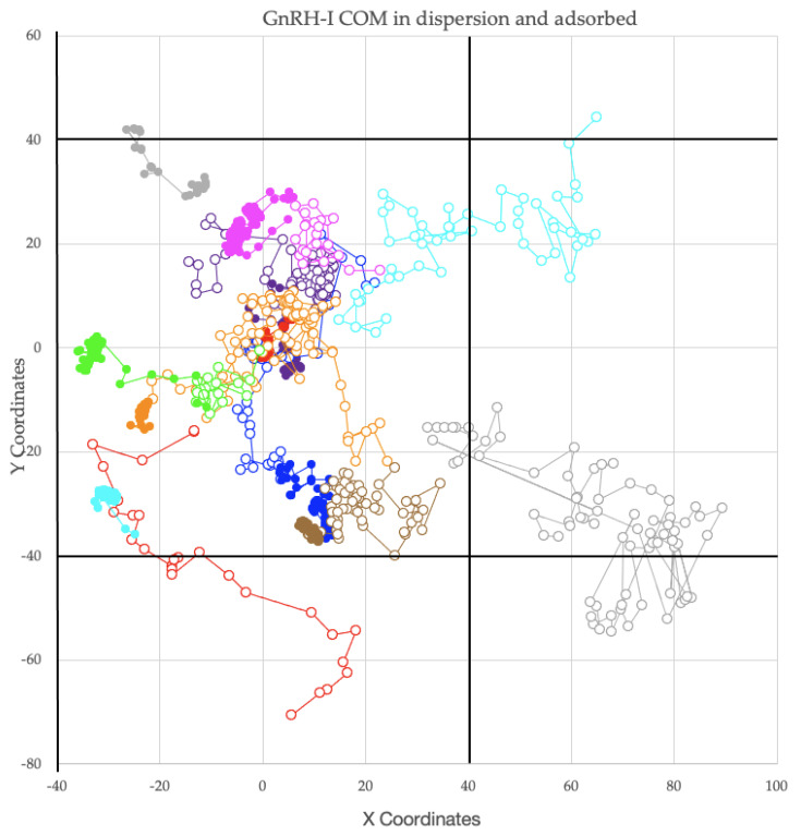
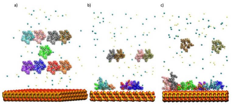
Molecular dynamics (MD) simulations can provide a detailed view of molecule behaviour at an atomic level, which can be useful when attempting to interpret experiments or design new systems. The decapeptide gonadotrophin-releasing hormone I (GnRH-I) is known to control fertility in mammals for both sexes. It was previously shown that inoculation with silica nanoparticles (SiNPs) coated with GnRH-I makes an effective anti-fertility vaccine due to how the peptide adsorbs to the nanoparticle and is presented to the immune system. In this paper, we develop and employ a protocol to simulate the development of a GnRH-I peptide adlayer by allowing peptides to diffuse and adsorb in a staged series of trajectories. The peptides start the simulation in an immobile state in solution above the model silica surface, and are then released sequentially. This facile approach allows the adlayer to develop in a natural manner and appears to be quite versatile. We find that the GnRH-I adlayer tends to be sparse, with electrostatics dominating the interactions. The peptides are collapsed to the surface and are seemingly free to interact with additional solutes, supporting the interpretations of the GNRH-I/SiNP vaccine system.

Keywords: GnRH-I; adsorption; molecular dynamics.


Conflict of interest statement

The authors declare no conflict of interests.

Figures

Figure 1
Figure 1
Simulated GnRH-I structure in solution using a transparent surface overlapped on the liquorice representation in VMD. Positive side chains are shown in blue, negative in red, and neutral in grey, and Arg8 is annotated.
Figure 2
Figure 2
The final configuration of the 9 GnRH-I peptides adsorbed to the model silica surface as viewed from above. The GnRH-I proteins are shown as transparent surfaces overlapped on opaque licorice VMD representations with each peptide given a different colour for ease of identification (see Table 1), while the silica as a yellow surface. The water molecules and ions are not shown for clarity.
Figure 3
Figure 3
The GnRH-I centre-of-mass (COM) z-coordinates measured for a period of 100 ns following their release. The trace colours match those given in Table 1.
Figure 4
Figure 4
Peptide COM (x, y) coordinate traces measured in Å. The traces are shown as continuous lines utilizing the periodic boundary conditions. Each peptide’s trace is coloured as in Table 1. Empty circles represent peptides before adsorption, solid circles after. The simulation cell is indicated with black solid lines, also indicating the periodic boundary conditions in the (x, y) directions.
Figure 5
Figure 5
GnRH-I COM distance from the silica surface as a function time, starting from when all 9 peptides were adsorbed. The peptide traces are coloured as in Table 1.
Figure 6
Figure 6
The simulation cell illustrating the GnRH-I starting positions above the model silica surface. (a) The initial 9 peptides; (b) the second simulation with 4 more peptides; (c) the third simulation with a further 2 peptides. GnRH-I proteins are displayed using a transparent surface overlapped on the VMD ‘liquorice’ representation, with each peptide given a different colour for ease of identification (see Table 1). Ions are shown as VdW spheres, Na+ ions are yellow while Cl ions are blue. The silica slab is represented by a surface with red oxygen and yellow silicon atoms. The water is not shown for clarity.

Similar articles

Cited by

References

    1. Yoshino R., Yasuo N., Sekijima M. Molecular Dynamics Simulation reveals the mechanism by which the Influenza Cap-dependent Endonuclease acquires resistance against Baloxavir marboxil. Sci. Rep. 2019;9:17464. doi: 10.1038/s41598-019-53945-1. - DOI - PMC - PubMed
    1. Tamamis P., López de Victoria A., Gorham R., Bellows-Peterson M., Pierou P., Floudas C., Morikis D., Archontis G. Molecular Dynamics in Drug Design: New Generations of Compstatin Analogs. Chem. Biol. Drug Des. 2012;79:703–718. doi: 10.1111/j.1747-0285.2012.01324.x. - DOI - PMC - PubMed
    1. Rallapalli K., Komor A., Paesani F. Computer simulations explain mutation-induced effects on the DNA editing by adenine base editors. Sci. Adv. 2020;6:eaaz2309. doi: 10.1126/sciadv.aaz2309. - DOI - PMC - PubMed
    1. Duan L., Guo X., Cong Y., Feng G., Li Y., Zhang J. Accelerated Molecular Dynamics Simulation for Helical Proteins Folding in Explicit Water. Front. Chem. 2019;7:540. doi: 10.3389/fchem.2019.00540. - DOI - PMC - PubMed
    1. Yoo J., Winogradoff D., Aksimentiev A. Molecular dynamics simulations of DNA–DNA and DNA–protein interactions. Curr. Opin. Struct. Biol. 2020;64:88–96. doi: 10.1016/j.sbi.2020.06.007. - DOI - PubMed

MeSH terms

LinkOut - more resources