

Genetic Patterns Found in the Nuclear Localization Signals (NLSs) Associated with EBV-1 and EBV-2 Provide New Insights into Their Contribution to Different Cell-Type Specificities
- PMID: 34073836
- PMCID: PMC8197229
- DOI: 10.3390/cancers13112569
Genetic Patterns Found in the Nuclear Localization Signals (NLSs) Associated with EBV-1 and EBV-2 Provide New Insights into Their Contribution to Different Cell-Type Specificities
Abstract
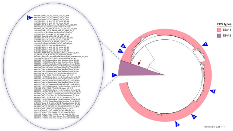
The Epstein-Barr virus (EBV) is a globally dispersed pathogen involved in several human cancers of B-cell and non-B-cell origin. EBV has been classified into EBV-1 and EBV-2, which have differences in their transformative ability. EBV-1 can transform B-cells into LCL more efficiently than EBV-2, and EBV-2 preferentially infects T-cell lymphocytes. The EBNA3A oncoprotein is a transcriptional regulator of virus and host cell genes, and is required in order to transform B-cells. EBNA3A has six peptide motifs called nuclear localization signals (NLSs) that ensure nucleocytoplasmic protein trafficking. The presence of multiple NLSs has been suggested to enhance EBNA3 function or different specificities in different cell types. However, studies about the NLS variability associated with EBV types are scarce. Based on a systematic sequence analysis considering more than a thousand EBNA3A sequences of EBV from different human clinical manifestations and geographic locations, we found differences in NLSs' nucleotide structures among EBV types. Compared with the EBNA3A EBV-1, EBNA3A EBV-2 has two of the six NLSs altered, and these mutations were possibly acquired by recombination. These genetic patterns in the NLSs associated with EBV-1 and EBV-2 provide new information about the traits of EBNA3A in EBV biology.
Keywords: EBV classification; EBV nuclear antigen EBNA 3A (EBNA3A); Epstein–Barr virus (EBV); nuclear localization signal (NLS); phylogeny; recombination.
Conflict of interest statement
The authors declare no conflict of interest.
Figures


Similar articles
-
Epstein-Barr Virus Nuclear Antigen 3 (EBNA3) Proteins Regulate EBNA2 Binding to Distinct RBPJ Genomic Sites.J Virol. 2015 Dec 30;90(6):2906-19. doi: 10.1128/JVI.02737-15. J Virol. 2015. PMID: 26719268 Free PMC article.
-
Epstein-Barr Virus (EBV) Latent Protein EBNA3A Directly Targets and Silences the STK39 Gene in B Cells Infected by EBV.J Virol. 2018 Mar 14;92(7):e01918-17. doi: 10.1128/JVI.01918-17. Print 2018 Apr 1. J Virol. 2018. PMID: 29367247 Free PMC article.
-
Epstein-Barr virus nuclear antigen 3A partially coincides with EBNA3C genome-wide and is tethered to DNA through BATF complexes.Proc Natl Acad Sci U S A. 2015 Jan 13;112(2):554-9. doi: 10.1073/pnas.1422580112. Epub 2014 Dec 24. Proc Natl Acad Sci U S A. 2015. PMID: 25540416 Free PMC article.
-
The Cooperative Functions of the EBNA3 Proteins Are Central to EBV Persistence and Latency.Pathogens. 2018 Mar 17;7(1):31. doi: 10.3390/pathogens7010031. Pathogens. 2018. PMID: 29562595 Free PMC article. Review.
-
The EBNA3 Family: Two Oncoproteins and a Tumour Suppressor that Are Central to the Biology of EBV in B Cells.Curr Top Microbiol Immunol. 2015;391:61-117. doi: 10.1007/978-3-319-22834-1_3. Curr Top Microbiol Immunol. 2015. PMID: 26428372 Review.
Cited by
-
Worldwide Prevalence of Epstein-Barr Virus in Patients with Burkitt Lymphoma: A Systematic Review and Meta-Analysis.Diagnostics (Basel). 2023 Jun 15;13(12):2068. doi: 10.3390/diagnostics13122068. Diagnostics (Basel). 2023. PMID: 37370963 Free PMC article. Review.
-
Epstein-Barr Virus-Associated Malignancies and Immune Escape: The Role of the Tumor Microenvironment and Tumor Cell Evasion Strategies.Cancers (Basel). 2021 Oct 16;13(20):5189. doi: 10.3390/cancers13205189. Cancers (Basel). 2021. PMID: 34680337 Free PMC article. Review.
-
Epstein-Barr Virus Infection in Cancer.Cancers (Basel). 2023 Sep 21;15(18):4659. doi: 10.3390/cancers15184659. Cancers (Basel). 2023. PMID: 37760627 Free PMC article.
References
-
- Corvalan A., Koriyama C., Akiba S., Eizuru Y., Backhouse C., Palma M., Argandoña J., Tokunaga M. Epstein-Barr virus in gastric carcinoma is associated with location in the cardia and with a diffuse histology: A study in one area of Chile. Int. J. Cancer. 2001;94:527–530. doi: 10.1002/ijc.1510. - DOI - PubMed
Grants and funding
- 3210687/Fondo Nacional de Desarrollo Científico y Tecnológico
- 3170826/Fondo Nacional de Desarrollo Científico y Tecnológico
- 3210618/Fondo Nacional de Desarrollo Científico y Tecnológico
- 3210604/Fondo Nacional de Desarrollo Científico y Tecnológico
- 11150622/Fondo Nacional de Desarrollo Científico y Tecnológico
- 1210440/Fondo Nacional de Desarrollo Científico y Tecnológico
- VRIP20P002/Universidad de La Frontera
- 21201835/Comisión Nacional de Investigación Científica y Tecnológica
- DIM20-0019/Universidad de La Frontera
- DI20-0160/Universidad de La Frontera
- DI20-0128/Universidad de La Frontera
- DIUA 226-2021/Universidad Autonoma de Chile
LinkOut - more resources
Full Text Sources
