

Radiomic Based Machine Learning Performance for a Three Class Problem in Neuro-Oncology: Time to Test the Waters?
- PMID: 34073840
- PMCID: PMC8197204
- DOI: 10.3390/cancers13112568
Radiomic Based Machine Learning Performance for a Three Class Problem in Neuro-Oncology: Time to Test the Waters?
Abstract
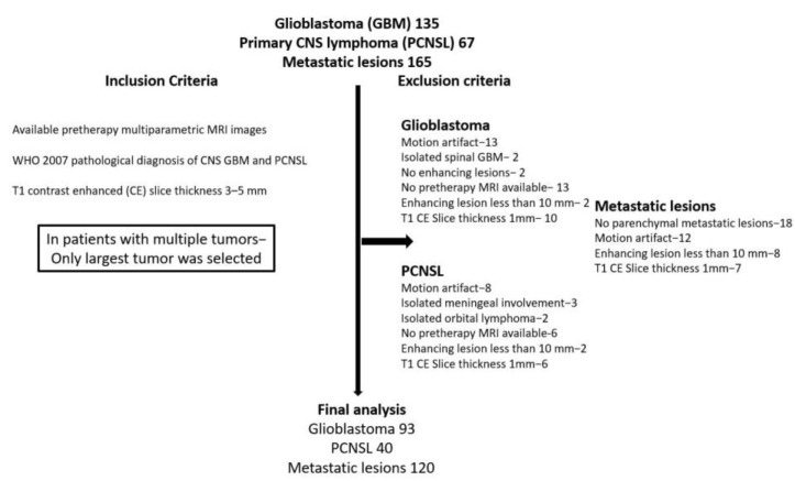
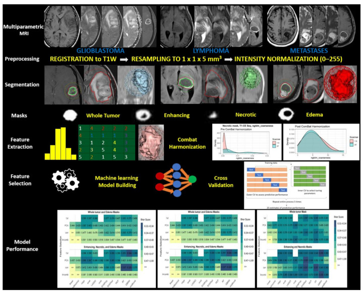
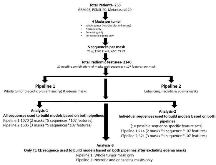
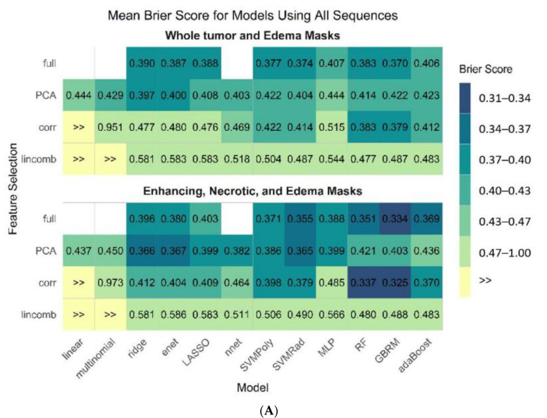
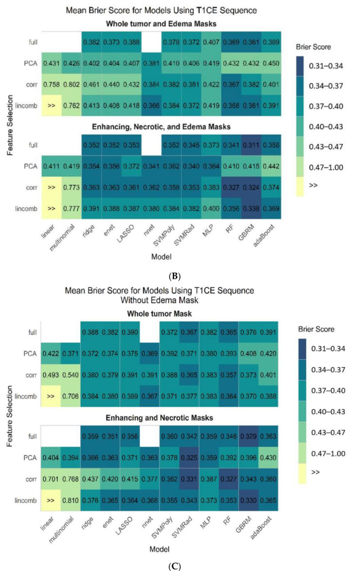
Prior radiomics studies have focused on two-class brain tumor classification, which limits generalizability. The performance of radiomics in differentiating the three most common malignant brain tumors (glioblastoma (GBM), primary central nervous system lymphoma (PCNSL), and metastatic disease) is assessed; factors affecting the model performance and usefulness of a single sequence versus multiparametric MRI (MP-MRI) remain largely unaddressed. This retrospective study included 253 patients (120 metastatic (lung and brain), 40 PCNSL, and 93 GBM). Radiomic features were extracted for whole a tumor mask (enhancing plus necrotic) and an edema mask (first pipeline), as well as for separate enhancing and necrotic and edema masks (second pipeline). Model performance was evaluated using MP-MRI, individual sequences, and the T1 contrast enhanced (T1-CE) sequence without the edema mask across 45 model/feature selection combinations. The second pipeline showed significantly high performance across all combinations (Brier score: 0.311-0.325). GBRM fit using the full feature set from the T1-CE sequence was the best model. The majority of the top models were built using a full feature set and inbuilt feature selection. No significant difference was seen between the top-performing models for MP-MRI (AUC 0.910) and T1-CE sequence with (AUC 0.908) and without edema masks (AUC 0.894). T1-CE is the single best sequence with comparable performance to that of multiparametric MRI (MP-MRI). Model performance varies based on tumor subregion and the combination of model/feature selection methods.
Keywords: CNS lymphoma; MRI; glioblastoma; machine learning; metastases; radiomics; texture.
Conflict of interest statement
Girish Bathla has research grants from Siemens AG, Forchheim, Germany, and the American Cancer Society, which are unrelated to the submitted work. The other authors report no associations that could be construed as conflict of interest.
Figures
References
-
- Neska-Matuszewska M., Bladowska J., Sąsiadek M., Zimny A. Differentiation of glioblastoma multiforme, metastases and primary central nervous system lymphomas using multiparametric perfusion and diffusion MR imaging of a tumor core and a peritumoral zone-Searching for a practical approach. PLoS ONE. 2018;13:e0191341. doi: 10.1371/journal.pone.0191341. - DOI - PMC - PubMed
-
- Swinburne N.C., Schefflein J., Sakai Y., Oermann E.K., Titano J.J., Chen I., Tadayon S., Aggarwal A., Doshi A., Nael K. Machine learning for semi-automated classification of glioblastoma, brain metastasis and central nervous system lymphoma using magnetic resonance advanced imaging. Ann. Transl. Med. 2019;7:232. doi: 10.21037/atm.2018.08.05. - DOI - PMC - PubMed
LinkOut - more resources
Full Text Sources
