Review
. 2021 May 24;14(11):2790.
doi: 10.3390/ma14112790.

Polymer Hernia Repair Materials: Adapting to Patient Needs and Surgical Techniques

Affiliations
Review

Polymer Hernia Repair Materials: Adapting to Patient Needs and Surgical Techniques

Marta Rodríguez et al. Materials (Basel). .

Abstract

Biomaterials and their applications are perhaps among the most dynamic areas of research within the field of biomedicine. Any advance in this topic translates to an improved quality of life for recipient patients. One application of a biomaterial is the repair of an abdominal wall defect whether congenital or acquired. In the great majority of cases requiring surgery, the defect takes the form of a hernia. Over the past few years, biomaterials designed with this purpose in mind have been gradually evolving in parallel with new developments in the different surgical techniques. In consequence, the classic polymer prosthetic materials have been the starting point for structural modifications or new prototypes that have always strived to accommodate patients' needs. This evolving process has pursued both improvements in the wound repair process depending on the implant interface in the host and in the material's mechanical properties at the repair site. This last factor is important considering that this site-the abdominal wall-is a dynamic structure subjected to considerable mechanical demands. This review aims to provide a narrative overview of the different biomaterials that have been gradually introduced over the years, along with their modifications as new surgical techniques have unfolded.

Keywords: Polypropylene; meshes; polytetrafluoroethylene.


Conflict of interest statement

The authors declare no conflict of interest.

Figures
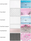
Figure 1
Figure 1
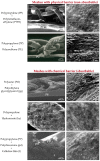
Macroscopic images of the different modifications in polytetrafluoroethylene (PTFE) meshes (left) and host tissue incorporation once implanted (right). Both surfaces, subcutaneous and peritoneal sides of the PTFE implants (Soft tissue Patch®, Dual mesh®, Dual Mesh Corduroy®-Dual Mesh Plus®, 30, 14, and 90 days post-implant, respectively, 100×) were encapsulated by host connective tissue. Scar tissue surrounds the PTFE implants, and some cells could be seen into the prosthetic interstices, at the inner third of the PTFE. Furthermore, in Mycro Mesh®, host tissue penetrates through the material micropores (60 days post-implant, 100×). Infinit Mesh® behaviour was similar to reticular meshes integration, like polypropylene, with connective tissue surrounding the mesh filaments (14 days post-implant, 100×). Scale bar: 100 µm.
Figure 2
Figure 2
(a) Diagram and (b) light microscopy (90 days post-implant, 100×) images showing tissue incorporation (cross-section), in PTFE meshes, once implanted in the abdominal wall. Meshes are encapsulated by vascularized connective tissue arranged as fibrous bundles running parallel to the prosthetic surface. Cells are observed inside the biomaterial, although they fail to penetrate beyond the outer third of the laminar sheet. Scale bar: 100 µm. (c) Scanning electron microscopy view of mesothelial covering (14 days, 500×). Scale bar: 20 µm.
Figure 3
Figure 3
(a) Diagrams and (b) light microscopy images (100×) showing tissue incorporation (cross-section) in polypropylene meshes, once implanted in the abdominal wall (90 days post-implant). The prosthetic filaments are surrounded by scar tissue in which the collagen fibres concentrically lay around the mesh filaments. The spaces between filaments are also occupied by scar tissue. Scale bar: 100 µm. (c) Scanning electron microscopy view of mesothelial covering (14 days, 500×). Scale bar: 20 µm.
Figure 4
Figure 4
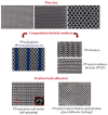
Macroscopic images of the different modifications to polypropylene meshes. Pore size: modifications in pore size have the objectives of minimizing minimize the quantity of foreign material in the host tissue, and improve the foreign body reaction and fibrosis without compromising mechanical resistance. Composition: hybrid meshes combine different components knitted or woven together to obtain a single mesh structure. Some of them incorporate an absorbable component (polyglecaprone-25 or polyglactin) to diminish the fibrosis reaction and amount of foreign material left in the body. Others include titanium or polymers like polyvinylidene fluoride. Fixation: self-adhesive meshes strive to achieve atraumatic mesh fixation (red box: polylactic acid hooks, scanning electron microscopy, 16×).
Figure 5
Figure 5
Composites consist of the combination of two different components linked together by suturing, heat-sealing, vacuum pressing or polymer adhesion.
Figure 6
Figure 6
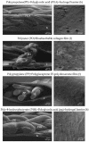
Scanning electron microscopy images of classic composites meshes (left) (lateral view, 50×. Scale bar: 500 µm) and mesothelial layer at the peritoneal side after implant (right, 500×. Scale bar: 20 µm). Composites include laminar components as adhesion barriers of physical (non absorbable) or chemical (absorbable) nature.
Figure 7
Figure 7
Scanning electron microscopy images of last generation composites (left) (lateral view, 50×. Scale bar: 500 µm) and mesothelial layer at the peritoneal side after implant (right, 500×. Scale bar: 20 µm). Nowadays, there is a tendency towards the use of reticular absorbable or partially absorbable components, and a short-term (14–30 days post-implant) absorbable laminar structure as adhesion barrier.
Figure 8
Figure 8
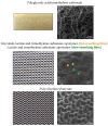
Research in new synthetic meshes are giving rise to fully absorbable products, like biocompatible synthetic polymers that are gradually absorbed by the host (macroscopic -left- and scanning electron microscopy images-right, 50×). Scale bar: 500 µm.

Similar articles

Cited by

References

    1. Ratner B.D., Hoffman A.S. Biomaterials Science: An Introduction to Materials in Medicine. 3rd ed. Academic Press; San Diego, CA, USA: 2012.
    1. Kingsnorth A., LeBlanc K. Hernias: Inguinal and incisional. Lancet. 2003;362:1561–1571. doi: 10.1016/S0140-6736(03)14746-0. - DOI - PubMed
    1. Luijendijk R.W., Hop W.C., van del Tol M.P., de Lange D.C., Braaksma M.M., Ijzermans J.N., Boelhouwer R.U., de Vriers B.C., Salu M.K., Wereldsma J.C., et al. A comparison of suture repair with mesh repair for incisional hernia. N. Engl. J. Med. 2000;343:392–398. doi: 10.1056/NEJM200008103430603. - DOI - PubMed
    1. Langer C., Liersch T., Kley C., Flosman M., Süss M., Siemer A. Twenty-five years of experience in incisional hernia surgery. A comparative retrospective study of 432 incisional hernia repairs. Der Chir. Z. Fur Alle Geb. Der Oper. Medizen. 2003;74:638–645. - PubMed
    1. Yang F. Radical tumor excision and immediate abdominal wall reconstruction in patients with aggressive neoplasm com-promised full thickness lower abdominal wall. Am. J. Surg. 2013;205:15–21. doi: 10.1016/j.amjsurg.2012.04.007. - DOI - PubMed

LinkOut - more resources