

Imaging Techniques for the Study of Fibrosis in Atrial Fibrillation Ablation: From Molecular Mechanisms to Therapeutical Perspectives
- PMID: 34073969
- PMCID: PMC8197293
- DOI: 10.3390/jcm10112277
Imaging Techniques for the Study of Fibrosis in Atrial Fibrillation Ablation: From Molecular Mechanisms to Therapeutical Perspectives
Abstract
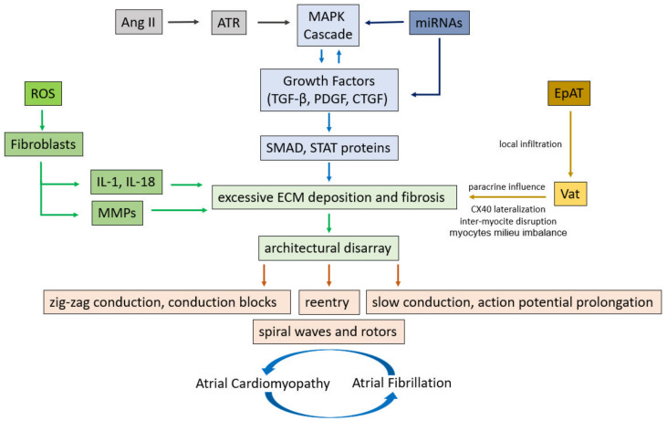
Atrial fibrillation (AF) is the most prevalent form of cardiac arrhythmia. It is often related to diverse pathological conditions affecting the atria and leading to remodeling processes including collagen accumulation, fatty infiltration, and amyloid deposition. All these events generate atrial fibrosis, which contribute to beget AF. In this scenario, cardiac imaging appears as a promising noninvasive tool for monitoring the presence and degree of LA fibrosis and remodeling. The aim of this review is to comprehensively examine the bench mechanisms of atrial fibrosis moving, then to describe the principal imaging techniques that characterize it, such as cardiac magnetic resonance (CMR) and multidetector cardiac computed tomography (MDCT), in order to tailor atrial fibrillation ablation to each individual.
Keywords: atrial cardiomyopathies; atrial failure; atrial fibrillation; atrial fibrosis; atrial remodeling; cardiac magnetic resonance; left atrial strain; multi detector computed tomography.
Conflict of interest statement
The authors declare no conflict of interest.
Figures
Similar articles
-
Assessment of left atrial volume and function in patients with permanent atrial fibrillation: comparison of cardiac magnetic resonance imaging, 320-slice multi-detector computed tomography, and transthoracic echocardiography.Eur Heart J Cardiovasc Imaging. 2014 May;15(5):532-40. doi: 10.1093/ehjci/jet239. Epub 2013 Nov 17. Eur Heart J Cardiovasc Imaging. 2014. PMID: 24247925
-
Relationship Between Fibrosis Detected on Late Gadolinium-Enhanced Cardiac Magnetic Resonance and Re-Entrant Activity Assessed With Electrocardiographic Imaging in Human Persistent Atrial Fibrillation.JACC Clin Electrophysiol. 2018 Jan;4(1):17-29. doi: 10.1016/j.jacep.2017.07.019. Epub 2017 Nov 6. JACC Clin Electrophysiol. 2018. PMID: 29479568 Free PMC article.
-
The association of left atrial low-voltage regions on electroanatomic mapping with low attenuation regions on cardiac computed tomography perfusion imaging in patients with atrial fibrillation.Heart Rhythm. 2015 May;12(5):857-64. doi: 10.1016/j.hrthm.2015.01.015. Epub 2015 Jan 13. Heart Rhythm. 2015. PMID: 25595922 Free PMC article.
-
The Role of Atrial Structural Remodeling in Atrial Fibrillation Ablation:An Imaging Point of View for Predicting Recurrence.J Atr Fibrillation. 2012 Aug 20;5(2):509. doi: 10.4022/jafib.509. eCollection 2012 Aug-Sep. J Atr Fibrillation. 2012. PMID: 28496757 Free PMC article. Review.
-
New Insights Into the Use of Cardiac Magnetic Resonance Imaging to Guide Decision Making in Atrial Fibrillation Management.Can J Cardiol. 2018 Nov;34(11):1461-1470. doi: 10.1016/j.cjca.2018.07.007. Epub 2018 Jul 12. Can J Cardiol. 2018. PMID: 30297256 Review.
Cited by
-
The Atrium in Atrial Fibrillation - A Clinical Review on How to Manage Atrial Fibrotic Substrates.Front Cardiovasc Med. 2022 Jul 4;9:879984. doi: 10.3389/fcvm.2022.879984. eCollection 2022. Front Cardiovasc Med. 2022. PMID: 35859594 Free PMC article. Review.
-
Left atrial function and fibrosis in lifelong endurance athletes: a cardiac magnetic resonance imaging study.Int J Cardiovasc Imaging. 2025 Jul;41(7):1321-1330. doi: 10.1007/s10554-025-03416-8. Epub 2025 May 21. Int J Cardiovasc Imaging. 2025. PMID: 40397349 Free PMC article.
-
Left Atrial Strain Helps Identifying the Cardioembolic Risk in Transient Ischemic Attacks Patients with Silent Paroxysmal Atrial Fibrillation.Ther Clin Risk Manag. 2022 Mar 10;18:213-222. doi: 10.2147/TCRM.S359490. eCollection 2022. Ther Clin Risk Manag. 2022. PMID: 35299625 Free PMC article.
-
Endothelial activation, cell-cell interactions, and inflammatory pathways in postoperative atrial fibrillation following cardiac surgery.Biomed J. 2024 Nov 26;48(4):100821. doi: 10.1016/j.bj.2024.100821. Online ahead of print. Biomed J. 2024. PMID: 39603594 Free PMC article.
-
Atrial cardiomyopathy: from cell to bedside.ESC Heart Fail. 2022 Dec;9(6):3768-3784. doi: 10.1002/ehf2.14089. Epub 2022 Aug 3. ESC Heart Fail. 2022. PMID: 35920287 Free PMC article. Review.
References
-
- Chugh S.S., Havmoeller R., Narayanan K., Singh D., Rienstra M., Benjamin E.J., Gillum R.F., Kim Y.-H., McAnulty J.H., Jr., Zheng Z.-J., et al. Worldwide epidemiology of atrial fibrillation: A global burden of disease 2010 study. Circulation. 2014;129:837–847. doi: 10.1161/CIRCULATIONAHA.113.005119. - DOI - PMC - PubMed
-
- Krijthe B.P., Kunst A., Benjamin E.J., Lip G.Y.H., Franco O.H., Hofman A., Witteman J.C.M., Stricker B.H., Heeringaet J. Projections on the number of individuals with atrial fibrillation in the European Union, from 2000 to 2060. Eur. Heart, J. 2013;34:2746–2751. doi: 10.1093/eurheartj/eht280. - DOI - PMC - PubMed
-
- Schnabel R.B., Yin X., Gona P., Larson M.G., Beiser A.S., McManus D.D., Newton-Cheh C., Lubitz S.A., Magnani J.W., Ellinor P.T., et al. 50 year trends in atrial fibrillation prevalence, incidence, risk factors, and mortality in the Framingham Heart Study: A cohort study. Lancet. 2015;11:154–162. - PMC - PubMed
-
- Ruddox V., Sandven I., Munkhaugen J., Skattebu J., Edvardsen T., Otterstad J.E. Atrial fibrillation and the risk for myocardial infarction, all-cause mortality and heart failure: A systematic review and meta-analysis. Eur. J. Prev. Cardiol. 2017;24:1555–1566. doi: 10.1177/2047487317715769. - DOI - PMC - PubMed
Publication types
LinkOut - more resources
Full Text Sources
