Meta-Analysis
. 2021 Aug 1;30(15):758-772.
doi: 10.1089/scd.2021.0065. Epub 2021 Jul 5.

Wharton's Jelly Mesenchymal Stem Cell-Derived Extracellular Vesicles Reduce SARS-CoV2-Induced Inflammatory Cytokines Under High Glucose and Uremic Toxin Conditions

Affiliations
Meta-Analysis

Wharton's Jelly Mesenchymal Stem Cell-Derived Extracellular Vesicles Reduce SARS-CoV2-Induced Inflammatory Cytokines Under High Glucose and Uremic Toxin Conditions

Vuong Cat Khanh et al. Stem Cells Dev. .

Abstract

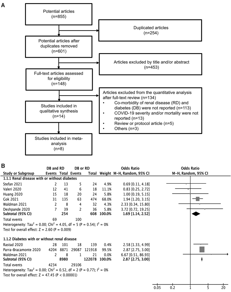
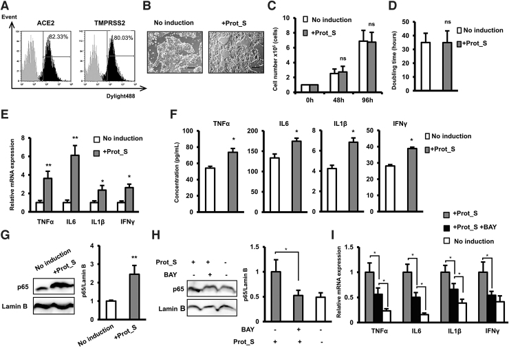
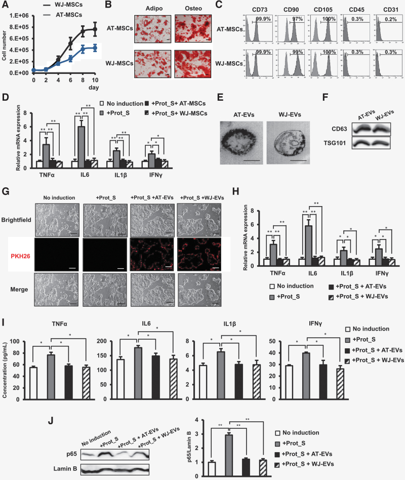
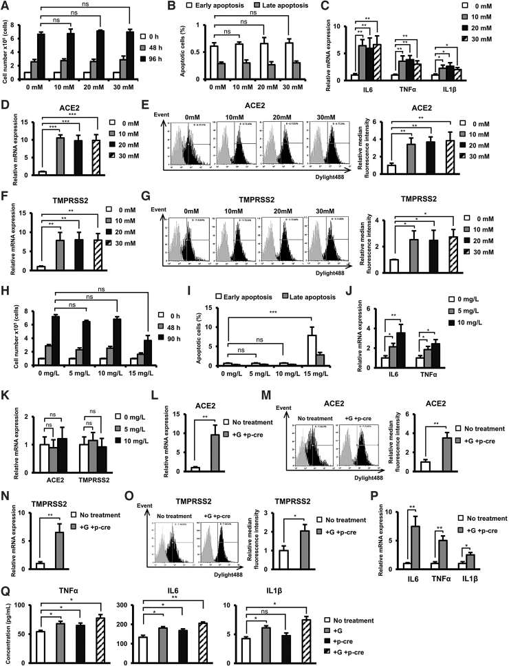
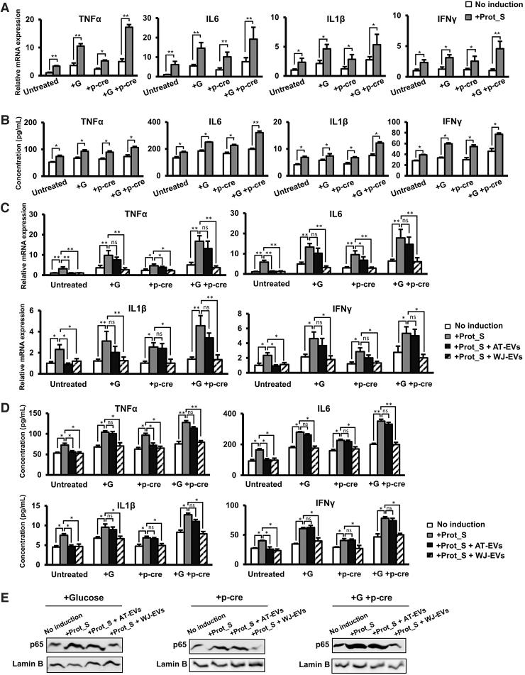
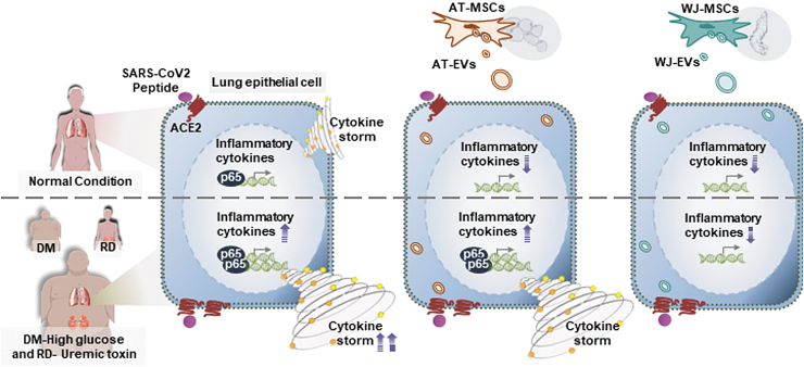
Cytokine storm is recognized as one of the factors contributing to organ failures and mortality in patients with COVID-19. Due to chronic inflammation, COVID-19 patients with diabetes mellitus (DM) or renal disease (RD) have more severe symptoms and higher mortality. However, the factors that contribute to severe outcomes of COVID-19 patients with DM and RD have received little attention. In an effort to investigate potential treatments for COVID-19, recent research has focused on the immunomodulation functions of mesenchymal stem cells (MSCs). In this study, the correlation between DM and RD and the severity of COVID-19 was examined by a combined approach with a meta-analysis and experimental research. The results of a systematic review and meta-analysis suggested that the odd of mortality in patients with both DM and RD was increased in comparison to those with a single comorbidity. In addition, in the experimental research, the data showed that high glucose and uremic toxins contributed to the induction of cytokine storm in human lung adenocarcinoma epithelial cells (Calu-3 cells) in response to SARS-CoV Peptide Pools. Of note, the incorporation of Wharton's jelly MSC-derived extracellular vesicles (WJ-EVs) into SARS-CoV peptide-induced Calu-3 resulted in a significant decrease in nuclear NF-κB p65 and the downregulation of the cytokine storm under high concentrations of glucose and uremic toxins. This clearly suggests the potential for WJ-EVs to reduce cytokine storm reactions in patients with both chronic inflammation diseases and viral infection.

Keywords: Calu-3 cells; DM; RD; SARS-CoV-2; cytokine storm; extracellular vesicles.


Conflict of interest statement

The authors declare no conflicts of interest in association with this study.

Figures

FIG. 1.
FIG. 1.
A systematic review and meta-analysis of the correlation between DM and RD and the risk of mortality in patients with COVID-19. (A) A flow diagram of the systematic review. A flow diagram of the number of articles identified and examined at each stage of the systematic review. A total of eight articles published from 2020 to 2021 met all inclusion criteria and were included in the meta-analysis. (B) Odds ratios for COVID-19-related mortality in patients with RD, DM, or RD and DM. DM, diabetes mellitus; RD, renal disease.
FIG. 2.
FIG. 2.
SARS-CoV-2 Peptide Pools induced cytokine storm in Calu-3 cells. (A) The surface expression of ACE2 and TMPRSS2 in Calu-3 cells examined by flow cytometry. (B) The morphology of Calu-3 cells under a microscope at 40 × magnification. Bars indicated 100 μm. (C) The proliferation of Calu-3 cells. (D) Doubling time of Calu-3 (n = 3). (E) The inflammatory cytokine gene expression in Calu-3 cells (n = 3). (F) The inflammatory cytokine secretion in Calu-3 cells (n = 4). (G) The nuclear p65 protein expression in Calu-3 cells. (H) The nuclear p65 protein expression in Calu-3 cells in the presence of NF-κB inhibitor BAY11-7082. I. The inflammatory cytokine gene expression in Calu-3 cells in the presence of NF-κB inhibitor. The data represent the mean ± SD. **P < 0.01, *P < 0.05, ns: no significance. The experiments were performed in triplicate.
FIG. 3.
FIG. 3.
MSC-EVs reduced the upregulation of the cytokine storm in Calu-3 cells induced by SARS-CoV-2 Peptide Pools. (A) Growth curve of MSCs. (B) Adipogenic and osteogenic differentiation of MSCs after 20 days. Adipo: Adipogenic differentiation, Osteo: Osteogenic differentiation. The photos were taken under a microscope at 200 × magnification. Bars indicated 100 μm. (C) Cell surface markers of MSCs. (D) The inflammatory cytokine expression in Calu-3 cell co-culture with MSCs. Four independent cell lines of MSCs were used in the above experiments (n = 4). (E) The morphology of EVs under a transmission electron microscopy. Bars indicated 100 nm. (F) The marker expression of EVs. (G) The incorporation of EVs into human lung epithelial cells after 24 h. The photos were taken under a microscope at 40 × magnification. Bars indicated 100 μm. (H) The inflammatory cytokine expression in Calu-3 cells incorporated with EVs. (I) The inflammatory cytokine secretion in Calu-3 cells incorporated with EVs (n = 4). (J) The nuclear p65 protein expression in Calu-3 cells incorporated with EVs. EVs isolated from four independent cell lines of MSCs were used in the above experiments (n = 4). The data represent the mean ± SD. **P < 0.01, *P < 0.05, ns: no significance. The experiments were performed in triplicate. MSC, mesenchymal stem cell; EV, extracellular vesicle.
FIG. 4.
FIG. 4.
High glucose or/and uremic toxin concentrations induced the expression of inflammatory cytokines in Calu-3 cells. (A) The proliferation of Calu-3 cells under high glucose concentrations. (B) The number of apoptotic cells under high glucose concentrations. (C) The gene expression of inflammatory cytokines in Calu-3 cells under high glucose concentrations. (D) The gene expression of ACE2 in Calu-3 cells under high glucose concentrations. Calu-3 was treated with 10, 20, and 30 mM glucose for 24 h. (E) The surface expression of ACE2 in Calu-3 cells under high glucose concentrations. (F) The gene expression of TMPRSS2 in Calu-3 cells under high glucose concentrations. (G) The surface expression of TMPRSS2 in Calu-3 cells under high glucose concentrations. (H) The proliferation of Calu-3 cells in the presence of p-cresol as an uremic toxin. (I) The number of apoptotic cells in the presence of p-cresol. (J) The gene expression of inflammatory cytokines in Calu-3 cells in the presence of p-cresol. (K) The gene expression of ACE2 in Calu-3 cells in the presence of p-cresol. Calu-3 cells were cultured under 5 or 10 mg/L p-cresol for 24 h. (L) The gene expression of ACE2 in Calu-3 cells in the presence of glucose and p-cresol. (M) The surface expression of ACE2 in Calu-3 cells in the presence of glucose and p-cresol. (N) The gene expression of TMPRSS2 in Calu-3 cells in the presence of glucose and p-cresol. (O) The surface expression of ACE2 in Calu-3 cells in the presence of glucose and p-cresol. (P) The gene expression of inflammatory cytokines in Calu-3 cells in the presence of glucose and p-cresol. Three independent experiments were performed for the above experiments (n = 3). (Q) The secretion of inflammatory cytokines in Calu-3 cells in the presence of glucose and p-cresol. (n = 4). Calu-3 cells were cultured under 30 mM Glucose and 10 mg/L p-cresol after 24 h. G: Glucose, p-cre: p-cresol. The data represent the mean ± SD. ***P < 0.001, **P < 0.01, *P < 0.05, ns: no significance. The experiments were performed in triplicate.
FIG. 5.
FIG. 5.
WJ-EVs reduced the cytokine storm of Calu-3 cells induced by SARS-CoV-2 Peptide Pools in high glucose and uremic toxin concentrations. (A) The inflammatory cytokine gene expression in Prot_S-induced Calu-3 cells (n = 3). (B) The inflammatory cytokine secretion in Prot_S-induced Calu-3 cells (n = 4). (C) The inflammatory cytokine gene expression in Prot_S-induced Calu-3 cells incorporated with EVs. (D) The inflammatory cytokine secretion in Prot_S-induced Calu-3 cells incorporated with EVs. (E) The nuclear p65 protein expression in Prot_S-induced Calu-3 cells. EVs isolated from four independent cell lines of MSCs were used in the above experiments (n = 4). G: Glucose, p-cre: p-cresol. The data represent the mean ± SD. **P < 0.01, *P < 0.05, ns: no significance. The experiments were performed in triplicate. WJ-EV, Wharton's jelly MSC-derived extracellular vesicle.
FIG. 6.
FIG. 6.
Proposed model: The ability of WJ-EVs in the downregulation of cytokine storm induced by SARS-CoV2 under high concentrations of glucose and uremic toxins. SARS-CoV-2 Peptide induces the cytokine storm, which might be involved in the activation of NF-κB p65 in human lung adenocarcinoma epithelial cells. In patients with DM and RD, the high concentrations of glucose and uremic toxins contribute to the severe cytokine storm induction by SARS-CoV2. WJ-EVs, but not AT-EVs, show the ability to reduce the NF-κB p65 and suppress the cytokine storm induced by SARS-CoV2 in the extraordinary worse situation as high glucose and uremic toxin level.

Similar articles

Cited by

References

    1. Shang J, Wan Y, Luo C, Ye G, Geng Q, Auerbach A and Li F. (2020). Cell entry mechanisms of SARS-CoV-2. Proc Natl Acad Sci USA 117:11727– 11734 - PMC - PubMed
    1. Merad M and Martin JC. (2020). Pathological inflammation in patients with COVID-19: a key role for monocytes and macrophages. Nat Rev Immunol 20:355–362 - PMC - PubMed
    1. Acharya D, Liu G and Gack MU. (2020). Dysregulation of type I interferon responses in COVID-19. Nat Rev Immunol 20:397–398 - PMC - PubMed
    1. Liu Q, Zhou Y and Yang Z. (2016). The cytokine storm of severe influenza and development of immunomodulatory therapy. Cell Mol Immunol 13:3–10 - PMC - PubMed
    1. Bhaskar S, Sinha A, Banach M, Mittoo S, Weissert R, Kass JS, Rajagopal S, Pai AR and Kutty S. (2020). Cytokine Storm in COVID-19-Immunopathological Mechanisms, Clinical Considerations, and Therapeutic Approaches: the REPROGRAM Consortium Position Paper. Front Immunol 11:1648. - PMC - PubMed

Publication types

MeSH terms

LinkOut - more resources