Review
. 2021 Nov 1;35(13):1093-1115.
doi: 10.1089/ars.2021.0018. Epub 2021 Jul 13.

ERO1-PDI Redox Signaling in Health and Disease

Affiliations
Review

ERO1-PDI Redox Signaling in Health and Disease

Vishwanath Jha et al. Antioxid Redox Signal. .

Abstract

Significance: Protein disulfide isomerase (PDI) and endoplasmic reticulum oxidoreductase 1 (ERO1) are crucial for oxidative protein folding in the endoplasmic reticulum (ER). These enzymes are frequently overexpressed and secreted, and they contribute to the pathology of neurodegenerative, cardiovascular, and metabolic diseases. Recent Advances: Tissue-specific knockout mouse models and pharmacologic inhibitors have been developed to advance our understanding of the cell-specific functions of PDI and ERO1. In addition to their roles in protecting cells from the unfolded protein response and oxidative stress, recent studies have revealed that PDI and ERO1 also function outside of the cells. Critical Issues: Despite the well-known contributions of PDI and ERO1 to specific disease pathology, the detailed molecular and cellular mechanisms underlying these activities remain to be elucidated. Further, although PDI and ERO1 inhibitors have been identified, the results from previous studies require careful evaluation, as many of these agents are not selective and may have significant cytotoxicity. Future Directions: The functions of PDI and ERO1 in the ER have been extensively studied. Additional studies will be required to define their functions outside the ER.

Keywords: ERO1; PDI; disease; inhibitors; oxidative ER stress; unfolded protein response.


Conflict of interest statement

Z.A. and K.L.O. are employees of Cayman Chemical Company Inc. The other authors declare no competing financial interests.

Figures

FIG. 1.
FIG. 1.
The binding site of FAD in ERO1α. (A, B) Glide docking was performed to verify the binding of FAD to ERO1α. Our docked model confirmed molecular interactions between FAD and ERO1α (in black) that were similar to those described in a previous report (3AHQ) (58) and identified new molecular interactions as shown in red. The abbreviation HIE255 represents His with hydrogen on nitrogen in the epsilon position. ER, endoplasmic reticulum; ERO1, ER oxidoreductase 1; FAD, flavin adenine dinucleotide. Color images are available online.
FIG. 2.
FIG. 2.
ERO1α-PDI redox cycling in the ER. In the highly oxidizing environment of the ER, unfolded proteins interact with the b′ domain of oxidized PDI and undergo oxidative protein folding. Misfolded substrate proteins can be reduced and refolded or isomerized to the appropriate native protein conformation. Reduced PDI is then reoxidized by ERO1α via direct hydrophobic interactions with its b′ domain and electron transfer from the a′ domain via a flexible Cys94 residue, resulting in the production of H2O2. ERO1β is expected to participate in similar redox cycling. ERO1α expression is upregulated by hypoxia, whereas ERO1β is expressed in response to the UPR. Mammals express several peroxidases, including peroxiredoxins (e.g., Prdx4) and GSH peroxidases (GPx7 and GPx8), to reduce H2O2-induced ER stress and can scavenge excess H2O2, thereby tightly regulating its concentration. Further, Prdx4 can utilize H2O2 to oxidize reduced PDI and restore the homeostatic redox state of ER. GPx7 and GPx8 also convert H2O2 to H2O. Oxidized GPx7 and GPx8 interact with and oxidize GRP78 (also known as BiP), thereby augmenting its chaperone activity. The increased levels of oxidized PDI promote the formation of the inhibitory disulfide bonds in ERO1α (Cys94–Cys131 and Cys99–Cys104) via which GSSG can induce oxidation of PDI. GPx7, GSH peroxidase 7; GRP78, glucose-regulated protein 78; GSH, glutathione; PDI, protein disulfide isomerase; UPR, unfolded protein response.
FIG. 3.
FIG. 3.
The role of PDI and ERO1α in neurodegenerative diseases. S-nitrosylation of PDI in association with nitrosative stress of neurons impairs its chaperone and enzymatic activities and induces the aggregation and accumulation of misfolded proteins (e.g., tau), thereby leading to neurotoxicity and various sequelae characteristic of AD and PD. The ER stress in brain cells results in upregulated expression of PDI and CHOP and may enhance PDI-ERO1α redox cycling and induce H2O2 overproduction and ER stress, thereby contributing to the pathology of HD. Further, mutations in the PDI b′ domain and PDI deletion may also be linked to the pathogenesis of ALS. AD, Alzheimer's disease; ALS, amyotrophic lateral sclerosis; C/EBP, CCAAT/enhancer-binding protein; CHOP, C/EBP-homologous protein; HD, Huntington's disease; PD, Parkinson's disease.
FIG. 4.
FIG. 4.
The role of ERO1α and PDI in cancer. The hypoxic tumor microenvironment induces the upregulation of ERO1α via HIF-1, suppresses T cell immunity, and promotes tumor angiogenesis, thereby facilitating metastasis and resulting in poor survival. The PDI expression may be upregulated by HIF-1α in gastric cancer cells. In glioma and breast cancer, upregulation of PDI may be a prognostic biomarker. Oxidized PDI increases PERK activity and the UPR in colorectal cancer. In AML, dysregulation of C/EBP-α results in aberrant myeloid differentiation. Upregulated levels of PDI may serve to suppress C/EBP function. AML, acute myeloid leukemia; HIF-1α, hypoxia-inducible factor-1α; PERK, protein kinase R-like endoplasmic reticulum kinase; VEGF, vascular endothelial growth factor.
FIG. 5.
FIG. 5.
The role of ERO1α and PDI in myocardial infarction. Hypoxia in cardiomyocytes induces expression of ERO1α and upregulation of the UPR. These responses enhance Ca2+ homeostasis and lead to progressive heart failure. Myocardial infarction results in the upregulated expression of PDI, thereby enhancing SOD1 activity and protection against cardiomyocyte apoptosis. SOD1, cytosolic superoxide dismutase 1.
FIG. 6.
FIG. 6.
The role of PDI and ERO1α in the progression of atherosclerosis. PDI and ERO1α function differently in the various cell types during the progression of atherosclerosis. PDI is upregulated in VSMCs, where it promotes ROS production through NOX1 or NOX4, which leads to cellular proliferation or apoptosis. In ECs, oxidized LDLs form 4-HNE-PDI, which impairs its activity. Hyperhomocysteinemia in hepatocytes induces ER stress, upregulates ER stress-related proteins (GRP78, PERK, ATF6, and XBP-1), and downregulates ERO1α expression. All of these pathways serve to aggravate the pathology associated with atherosclerosis. 4-HNE, 4-hydroxynonenal; ATF6, activating transcription factor 6; LDLs, low-density lipoproteins; NOX1, NADPH oxidase 1; ROS, reactive oxygen species; VSMC, vascular smooth muscle cell; XBP-1, X-box binding protein 1.
FIG. 7.
FIG. 7.
The role of PDI and ERO1α in ischemic stroke. CHOP and ERO1α are upregulated in the hippocampus, and PDI is upregulated in the cerebral cortex during ischemic stroke. Platelet-derived PDI binds to GPIbα and enhances its ligand-binding function, thereby contributing to platelet–neutrophil aggregation and vascular occlusion. GPIbα, glycoprotein Ibα.
FIG. 8.
FIG. 8.
The role of extracellular PDI in arterial and venous thrombosis. PDI is released from activated intravascular cells, including activated (adherent) platelets and ECs, in response to vascular injury. Extracellular PDI promotes the ligand-binding activity of GPIbα and αIIbβ3 by modifying their disulfide bonds, thereby inducing the formation of platelet thrombi. PDI also enhances TF activity, which leads to the generation of thrombin and fibrin. Both platelet aggregation and fibrin clots contribute to arterial/venous thrombosis and vascular occlusion. TF, tissue factor.
FIG. 9.
FIG. 9.
The role of ERO1α and PDI in diabetes. Insulin production is tightly regulated in the β cells of the pancreas and relies on the appropriate folding of proinsulin by ERO1β and PDI. Misfolded proinsulin leads to increased UPR and ER stress and, ultimately, β cell dysfunction and death, which characterizes the progression of diabetes. High glucose levels in adipocytes result in mitochondrial stress. PDI activity is reduced due to succinylation, which impairs the synthesis and secretion of the antidiabetic hormone, adiponectin. ERO1α also plays a crucial role in releasing adiponectin from ERp44.
FIG. 10.
FIG. 10.
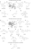
The structures of ERO1α inhibitors. (A) EN460, (B) QM295, and (C) Erodoxin.
FIG. 11.
FIG. 11.
The structures of PDI inhibitors and their binding site. (A–G) Inhibitors that bind to the b′ domain of PDI. (H–R) Inhibitors that bind to the active site(s) of PDI. (B, H) Glide docking was performed to confirm the binding site of isoquercetin and LOC14 to PDI. (S) No binding site has been reported for this inhibitor.

Similar articles

Cited by

References

    1. Abad Rico JI, Llau Pitarch JV, and Rocha E. Overview of venous thromboembolism. Drugs 70 Suppl 2: 3–10, 2010. - PubMed
    1. Abe JI, Ko KA, Kotla S, Wang Y, Paez-Mayorga J, Shin IJ, Imanishi M, Vu HT, Tao Y, Leiva-Juarez MM, Thomas TN, Medina JL, Won JH, Fujii Y, Giancursio CJ, McBeath E, Shin JH, Guzman L, Abe RJ, Taunton J, Mochizuki N, Faubion W, Cooke JP, Fujiwara K, Evans SE, and Le NT. MAGI1 as a link between endothelial activation and ER stress drives atherosclerosis. JCI Insight 4: e125570, 2019. - PMC - PubMed
    1. Alhammad R, Khunchai S, Tongmuang N, Limjindaporn T, Yenchitsomanus PT, Mutti L, Krstic-Demonacos M, and Demonacos C. Protein disulfide isomerase A1 regulates breast cancer cell immunorecognition in a manner dependent on redox state. Oncol Rep 44: 2406–2418, 2020. - PMC - PubMed
    1. Appenzeller-Herzog C, Riemer J, Christensen B, Sorensen ES, and Ellgaard L. A novel disulphide switch mechanism in Ero1alpha balances ER oxidation in human cells. EMBO J 27: 2977–2987, 2008. - PMC - PubMed
    1. Araki K, Iemura S, Kamiya Y, Ron D, Kato K, Natsume T, and Nagata K. Ero1-alpha and PDIs constitute a hierarchical electron transfer network of endoplasmic reticulum oxidoreductases. J Cell Biol 202: 861–874, 2013. - PMC - PubMed

Publication types

MeSH terms