. 2021 Jun 1;20(1):81.
doi: 10.1186/s12943-021-01375-x.

The circular RNA circSPARC enhances the migration and proliferation of colorectal cancer by regulating the JAK/STAT pathway

Affiliations

The circular RNA circSPARC enhances the migration and proliferation of colorectal cancer by regulating the JAK/STAT pathway

Jiaqi Wang et al. Mol Cancer. .

Abstract

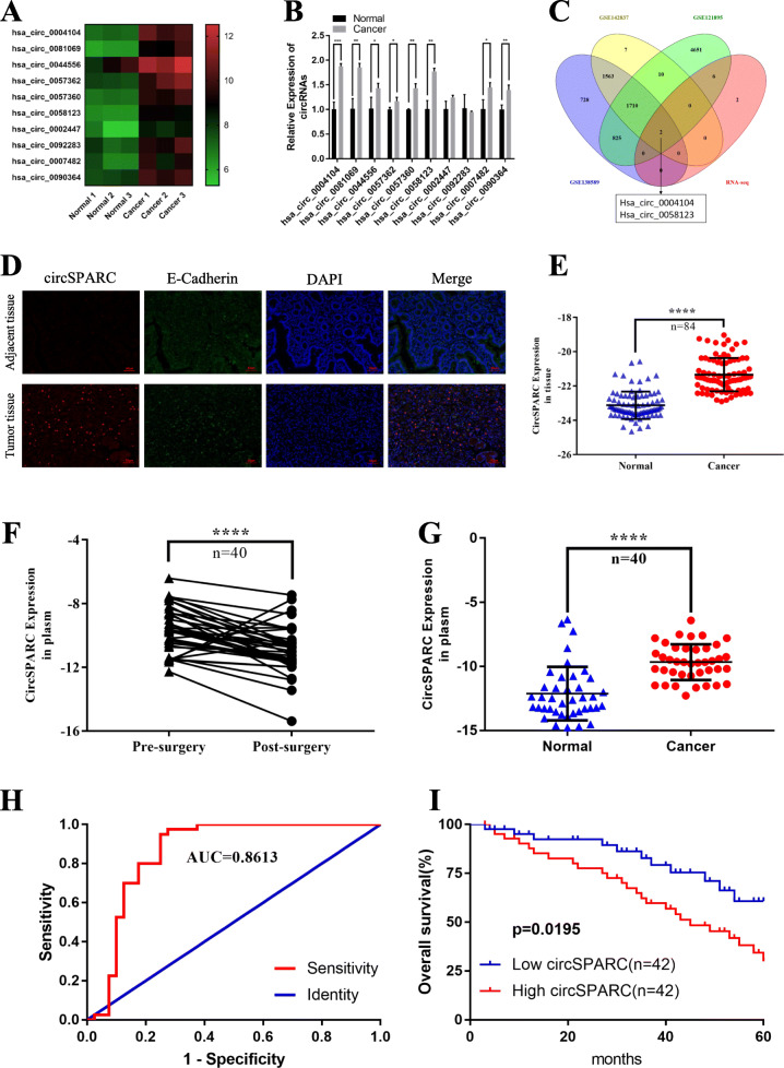
Background: Noncoding RNAs such as circular RNAs (circRNAs) are abundant in the human body and influence the occurrence and development of various diseases. However, the biological functions of circRNAs in colorectal cancer (CRC) are largely unknown.

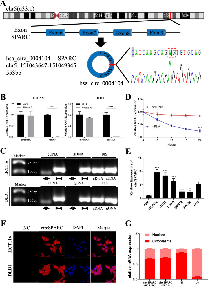
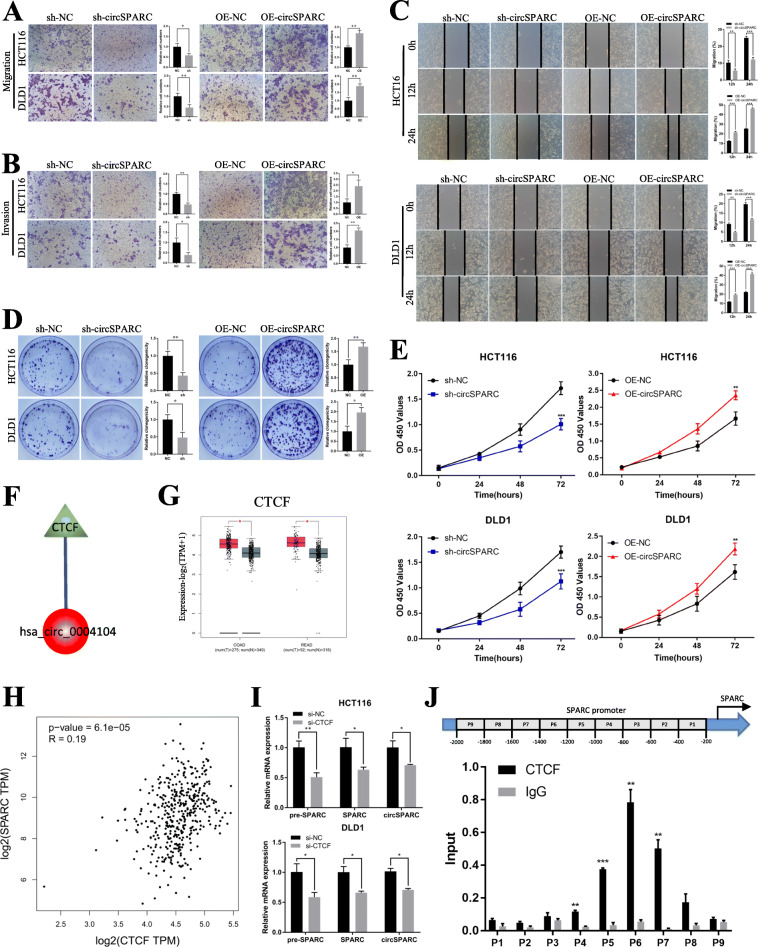
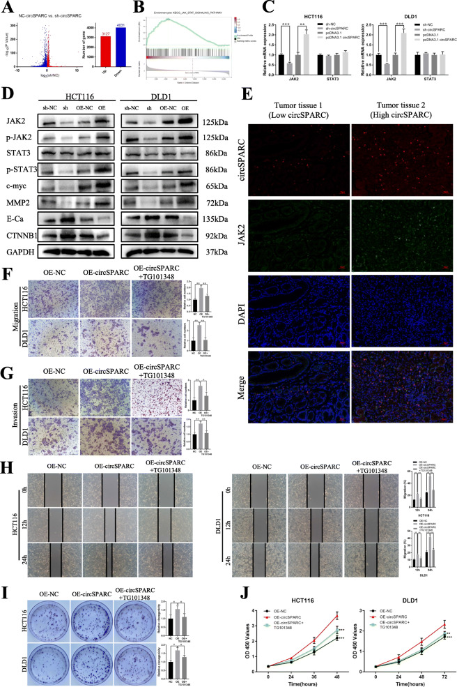
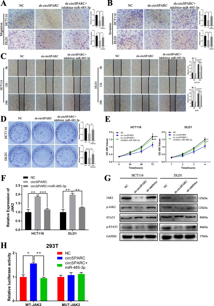
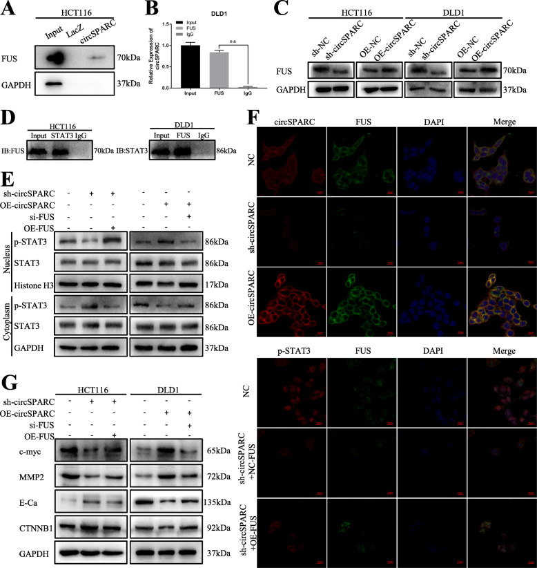
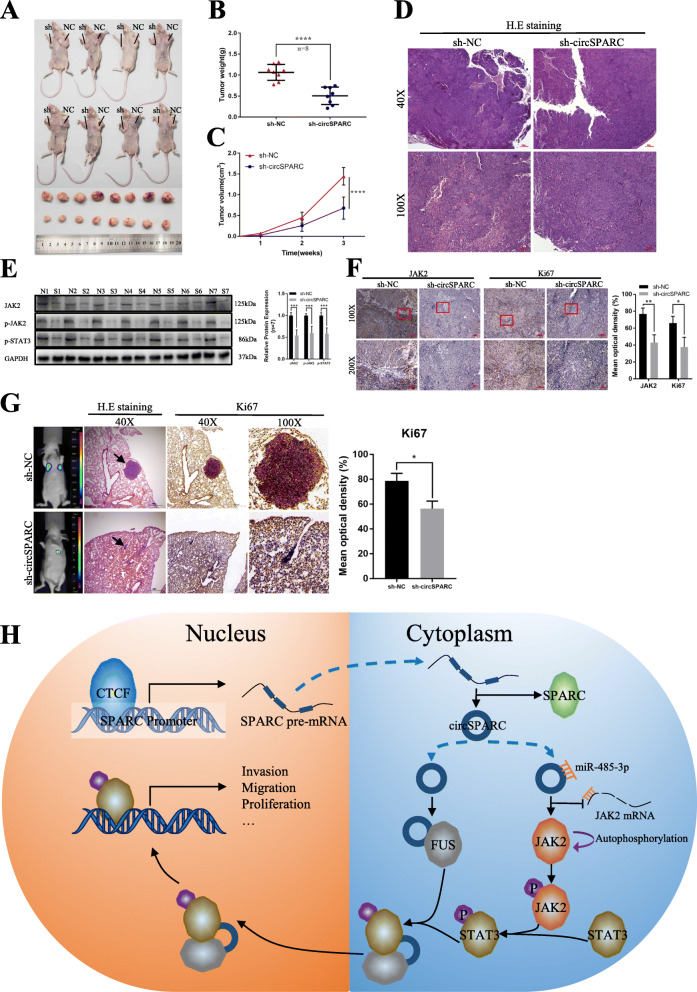
Methods: RT-qPCR was used to detect the expression of circRNAs and mRNA in CRC cells and tissues. Fluorescence in situ hybridization (FISH) was used to analyze the location of circSPARC. Function-based experiments were performed using circSPARC knockdown and overexpression cell lines in vitro and in vivo, including CCK8, colony formation, transwell and metastasis models. Mechanistically, luciferase reporter assay, western blots, RNA immunoprecipitation (RIP), Chromatin isolation by RNA purification (ChIRP) and immunohistochemical stainings were performed.

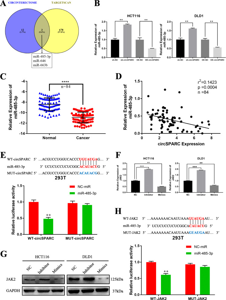
Results: CircSPARC was upregulated in both the tissues and plasma of CRC patients. High expression of circSPARC was associated with advanced TNM stage, lymph node metastases, and poor survival. Silencing circSPARC inhibited CRC cell migration and proliferation in vitro and vivo. Mechanistically, circSPARC sponged miR-485-3p to upregulate JAK2 expression and ultimately contribute to the accumulation of phosphorylated (p)-STAT3. Besides, circSPARC recruited FUS, which facilitated the nuclear translocation of p-STAT3.

Conclusions: These findings suggest that circSPARC might serve as a potential diagnostic and prognostic biomarker and a therapeutic target for CRC treatment by regulating JAK2/STAT3 pathway.

Keywords: Biomarker; Colorectal cancer; JAK/STAT signalling pathway; circRNA; circSPARC.


Conflict of interest statement

The authors declare that they have no conflicts of interest.

Figures

Fig. 1
Fig. 1
circSPARC was upregulated in CRC. A. Heatmap of RNA-Seq analysis of top 10 differentially high expressed circRNAs generated from three paired of CRC and corresponding adjacent non-tumoral tissues. Red in the heatmap denotes upregulation, green denotes downregulation. B. The expression of the top 10 differentially high expressed circRNAs were verified in CRC and corresponding adjacent non-tumoral tissues by RT-qPCR. C. The intersection of RNA-seq results and three data from the GEO database. D. The co-localization of epithelial marker E-Cadherin and circSPARC was applied by FISH-IF staining assay in CRC and corresponding adjacent non-tumoral tissues (200X). The results showed that circSPARC expressed higher in tumor tissues. E. RT-qPCR was utilized to analyze the circSPARC expression in 84 pairs of CRC tissues and corresponding adjacent non-tumoral tissues. F. Expression of circSPARC was detected by RT-qPCR in 40 pairs of pre-surgery and post-surgery patients plasm. G. RT-qPCR was applied to analyze the circSPARC expression in 40 pairs of CRC patients and general people plasm. H. ROC analysis of the prognostic sensitivity and specificity for CRC patients and general people detected by the expression of circSPARC in plasm. I. Kaplan-Meier overall survival curves according to circSPARC expression in our cohort. *P < 0.05, **P < 0.01, ***P < 0.001 and ****P < 0.0001
Fig. 2
Fig. 2
The characteristics of circSPARC. A. Schematic illustration indicating the generation of circSPARC from its host gene, and validation by Sanger sequencing. B. RT-qPCR for the abundance of circSPARC and its host gene mRNA in CRC cells treated with RNase R. C. PCR assay with divergent (◀▶) and convergent (▶◀) primers showing the amplification of circSPARC from cDNA or gDNA of CRC cell lines, while 18 s was used as a positive control. It can be observed that circSPARC was amplified in cDNA but not in gDNA while linear SPARC was amplified in both. D. Transcript half-life of circSPARC and its linear transcript in HCT116 cells treated with the transcription inhibitor actinomycin D. E. RT-qPCR indicated the circSPARC expressed higher in CRC cell lines (HCT116, DLD1, LoVo, SW480, SW620 and HT29) than in the normal colorectal epithelium cell line (FHC). F and G. FISH assay and nuclear-cytoplasm separation RT-qPCR data showed that circSPARC existed in both cytoplasm and nucleus, but primarily localized in the cytoplasm in CRC cells. *P < 0.05, **P < 0.01, ***P < 0.001 and ****P < 0.0001
Fig. 3
Fig. 3
circSPARC regulated the proliferation and invasion of CRC cells and CTCF may function as the upstream of circSPARC. A and B. The transwell assay was performed for invasion and migration of CRC cells. C. The wound healing assay was performed for migration of CRC cells. D. The colony formation assay was performed for proliferation of CRC cells. E. The CCK-8 assay was performed for proliferation of CRC cells. F. TRcirc tool predicted that CTCF can function as the potential upstream of circSPARC. G. GEPIA database exhibited that CTCF is upregulated in CRC. H. GEPIA database showed that CTCF has a positive correlation with SPARC in CRC. I. RT-qPCR results showed that the level of SPARC pre-mRNA, circSPARC and SPARC mRNA were all decreased when CTCF was knockdown in HCT116 and DLD1 cells. J. P1-P9 showed the regions of SPARC promoter detected by the paired primers. ChIP assay revealed that CTCF mainly binds at P5, P6 and P7. *P < 0.05, **P < 0.01, ***P < 0.001 and ****P < 0.0001
Fig. 4
Fig. 4
circSPARC regulated the JAK2/STAT3 signalling pathway. A and B. RNA-seq results showed that circSPARC had connection with JAK/STAT signalling pathway. C. RT-qPCR results showed that circSPARC had positive regulation on JAK2 but not STAT3. D. Western blot analysis showed that both silencing and overexpression circSPARC can regulate JAK2/STAT3 signalling pathway and its downstream genes expect STAT3. E. FISH-IF assay showed that JAK2 expressed higher in CRC tissues with high level of circSPARC. F and G. The transwell assay showed that the enhanced invasion and migration abilities were reversed by TG101348, an inhibitor of JAK2/STAT3 signalling. H. The wound healing assay showed that the enhanced migration abilities were reversed by TG101348, an inhibitor of JAK2/STAT3 signalling. I and J. The colony formation and CCK-8 assay showed that the enhanced proliferation abilities were reversed by TG101348. *P < 0.05, **P < 0.01, ***P < 0.001 and ****P < 0.0001
Fig. 5
Fig. 5
miR-485-3p was directly bound to circSPARC and suppresses JAK2 activity. A and B. miR-485-3p, miR-646 and miR-663b were predicted that they can bind to circSPARC by two databases and were verified in CRC cells. C. RT-qPCR was utilized to analyse the miR-485-3p expression in 84 pairs of CRC tissues and corresponding adjacent non-tumoral tissues. D. The negative interaction within miR-485-3p and circSPARC. E. Luciferase reporter assay functionally verified the interaction within the circSPARC and miR-485-3p in 293 T cell. F and G. RT-PCR and western blot analysis detected the JAK2 level with transfecting miR-485-3p mimics or inhibitor in CRC cell lines. H. Luciferase reporter assay functionally verified the interaction within the JAK2 and miR-485-3p in 293 T cell. *P < 0.05, **P < 0.01, ***P < 0.001 and ****P < 0.0001
Fig. 6
Fig. 6
circSPARC regulates JAK2 expression and inhibits CRC cells activities by targeting miR-485-3p. A and B. The transwell assay demonstrated that cell migration and invasion abilities of CRC cells transfected with sh-circSPARC were counteracted when co-transfected with miR-485-3p inhibitor. C. The wound healing assay demonstrated that cell migration abilities of CRC cells transfected with sh-circSPARC were counteracted when co-transfected with miR-485-3p inhibitor. D and E. The colony formation and CCK-8 assay demonstrated that cell proliferation abilities of CRC cells transfected with sh-circSPARC were counteracted when co-transfected with miR-485-3p inhibitor. F. RT-qPCR results showed that the level of JAK2 transfected with circSPARC plasmid was reversed when co-transfected with miR-485-3p mimics in CRC cells. G. Western blot analysis showed that the level of JAK2 and its downstream genes transfected with sh-circSPARC was rescued when co-transfected with miR-485-3p inhibitor in CRC cells. H. Luciferase reporter assay functionally showed that overexpression of circSPARC increased the activity while co-transfection of the miR-485-3p mimic could eliminate this effect on the wild-type JAK2 sequence in 293 T cell. *P < 0.05, **P < 0.01, ***P < 0.001 and ****P < 0.0001
Fig. 7
Fig. 7
circSPARC intensified the nuclear translocation of STAT3 in CRC via recruiting FUS. A and B. ChIRP and RIP assay were conducted to prove the interaction between circSPARC and FUS in HCT116 and DLD1 cells. C. Western blot assay verified the effect of circSPARC on FUS protein in CRC cells. D. The interaction between FUS and STAT3 was confirmed by coIP assay. E. Western blot assay was applied to detect the level of STAT3 after nuclear-cytoplasm separation in DLD1 cell. F. The co-localization of circSPARC and FUS, as well as that of FUS and STAT3, was validated by IF analysis. *P < 0.05, **P < 0.01, ***P < 0.001 and ****P < 0.0001
Fig. 8
Fig. 8
CircSPARC enhances tumour growth and metastasis in vivo. A-C. HCT116 cells stably transfected with sh-circSPARC or vector were injected subcutaneously into the BALB/c nude mice. Tumour volume and weight were dramatically decreased in sh-circSPARC group. D. H & E staining was applied to identify the subcutaneous tumours. E. The expression of JAK2 in tumours were analysed by western blot assay. N: sh-NC group. S: sh-circSPARC group. F. IHC assay demonstrated the level of JAK2 and Ki67 in pairs of tumours. G. After HCT116 cells with knockdown circSPARC injected into the tail vein of nude mice, in vivo fluorescence imaging, the gross lesion in lung tissues, H. E staining and the IHC of Ki67 of metastatic nodules in the lungs were observed. H. The hypothetical model depicts the roles of circSPARC in the promotion of CRC. *P < 0.05, **P < 0.01, ***P < 0.001 and ****P < 0.0001

Similar articles

Cited by

References

    1. Brody H. Colorectal cancer. Nature. 521(2015):S1. - PubMed
    1. Keum N, Giovannucci E. Global burden of colorectal cancer: emerging trends, risk factors and prevention strategies. Nat Rev Gastroenterol Hepatol. 2019;16(12):713–732. doi: 10.1038/s41575-019-0189-8. - DOI - PubMed
    1. Buccafusca G, Proserpio I, Tralongo AC, Rametta Giuliano S, Tralongo P. Early colorectal cancer: diagnosis, treatment and survivorship care. Crit Rev Oncol Hematol. 2019;136:20–30. doi: 10.1016/j.critrevonc.2019.01.023. - DOI - PubMed
    1. Tol J, Koopman M, Cats A, Rodenburg CJ, Creemers GJ, Schrama JG, Erdkamp FL, Vos AH, van Groeningen CJ, Sinnige HA, Richel DJ, Voest EE, Dijkstra JR, Vink-Börger ME, Antonini NF, Mol L, van Krieken JH, Dalesio O, Punt CJ. Chemotherapy, bevacizumab, and cetuximab in metastatic colorectal cancer. N Engl J Med. 2009;360(6):563–572. doi: 10.1056/NEJMoa0808268. - DOI - PubMed
    1. Dekker E, Tanis PJ, Vleugels JLA, Kasi PM, Wallace MB. Colorectal cancer. Lancet (London, England) 2019;394:1467–1480. doi: 10.1016/S0140-6736(19)32319-0. - DOI - PubMed

Publication types

MeSH terms