

Hypoxic tumor-derived exosomal miR-31-5p promotes lung adenocarcinoma metastasis by negatively regulating SATB2-reversed EMT and activating MEK/ERK signaling
- PMID: 34074322
- PMCID: PMC8167983
- DOI: 10.1186/s13046-021-01979-7
Hypoxic tumor-derived exosomal miR-31-5p promotes lung adenocarcinoma metastasis by negatively regulating SATB2-reversed EMT and activating MEK/ERK signaling
Abstract
Background: Exosomes have emerged as critical mediators of intercellular communication. Hypoxia is widely recognized as a key regulator of tumor aggressiveness, and significantly affects exosome release by tumor cells. However, the effects of exosomes derived from hypoxic lung adenocarcinoma (LUAD) cells are poorly understood.
Methods: Samples of miRNA isolated from hypoxic LUAD cell-derived exosomes (HExo) and normoxic LUAD cell-derived exosomes (NExo) were sequenced to identify miRNAs that might mediate tumor progression. Exosomal miRNA was co-cultured with LUAD cells to assess its biological effects on cell migration and metastasis both in vitro and in vivo. The cellular target of exosomal miRNA was confirmed by dual-luciferase assays. Western blot studies showed that exosomal miRNA regulated the related pathway. The availability of circulating exosomal miRNA derived from plasma was also evaluated.
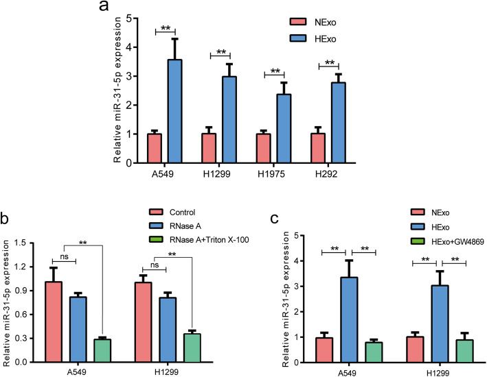
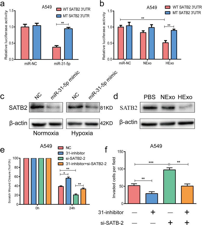
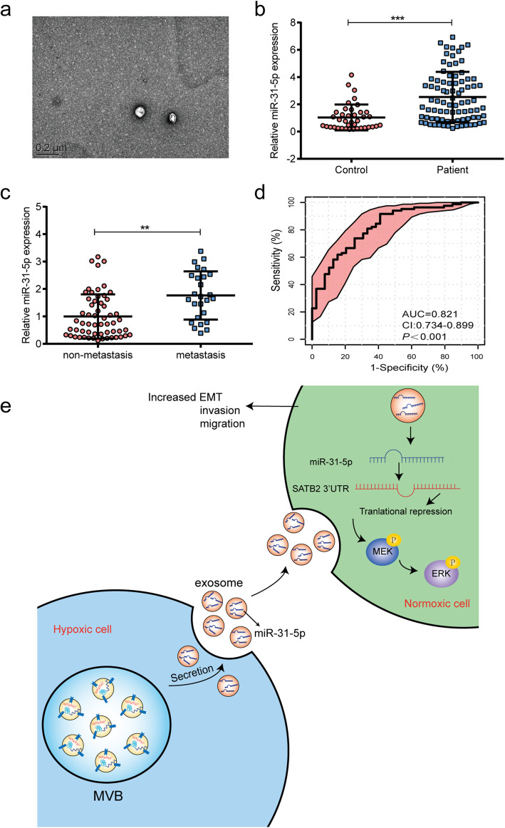
Results: We found that HExo could significantly enhance the migration and invasion of normoxic LUAD cells. MiRNA sequencing results suggested that miR-31-5p was largely internalized within HExo and could be taken up by normoxic LUAD cells. Exosomal miR-31-5p was found to directly target Special AT-Rich Sequence-Binding Protein 2 (SATB2)-revered epithelial mesenchymal transition and significantly increase activation of MEK/ERK signaling, thereby contributing to tumor progression both in vitro and in vivo. Furthermore, higher levels of circulating exosomal miR-31-5p were detected in LUAD patients, especially in patients with metastatic disease.
Conclusions: Our findings demonstrate that exosomal miR-31-5p exerts a crucial role in LUAD progression, and could serve as a diagnostic biomarker for LUAD.
Keywords: Lung adenocarcinoma; SATB2; exosome; hypoxia; miR-31-5p.
Conflict of interest statement
The authors declare that they have no competing interests.
Figures


Similar articles
-
miR-21-5p promotes lung adenocarcinoma cell proliferation, migration and invasion via targeting WWC2.Cancer Biomark. 2020;28(4):549-559. doi: 10.3233/CBM-201489. Cancer Biomark. 2020. PMID: 32623387
-
Exosomal miR-499a-5p promotes cell proliferation, migration and EMT via mTOR signaling pathway in lung adenocarcinoma.Exp Cell Res. 2019 Jun 15;379(2):203-213. doi: 10.1016/j.yexcr.2019.03.035. Epub 2019 Apr 10. Exp Cell Res. 2019. PMID: 30978341
-
LncRNA TTN-AS1 promotes migration, invasion, and epithelial mesenchymal transition of lung adenocarcinoma via sponging miR-142-5p to regulate CDK5.Cell Death Dis. 2019 Jul 30;10(8):573. doi: 10.1038/s41419-019-1811-y. Cell Death Dis. 2019. PMID: 31363080 Free PMC article.
-
The role of CAF derived exosomal microRNAs in the tumour microenvironment of melanoma.Biochim Biophys Acta Rev Cancer. 2021 Jan;1875(1):188456. doi: 10.1016/j.bbcan.2020.188456. Epub 2020 Oct 22. Biochim Biophys Acta Rev Cancer. 2021. PMID: 33153973 Review.
-
MicroRNA-200c in Cancer Generation, Invasion, and Metastasis.Int J Mol Sci. 2025 Jan 16;26(2):710. doi: 10.3390/ijms26020710. Int J Mol Sci. 2025. PMID: 39859424 Free PMC article. Review.
Cited by
-
HSD17B6 downregulation predicts poor prognosis and drives tumor progression via activating Akt signaling pathway in lung adenocarcinoma.Cell Death Discov. 2021 Nov 8;7(1):341. doi: 10.1038/s41420-021-00737-0. Cell Death Discov. 2021. PMID: 34750355 Free PMC article.
-
Recent advances on high-efficiency of microRNAs in different types of lung cancer: a comprehensive review.Cancer Cell Int. 2023 Nov 20;23(1):284. doi: 10.1186/s12935-023-03133-z. Cancer Cell Int. 2023. PMID: 37986065 Free PMC article. Review.
-
Identification of New Prognostic Genes and Construction of a Prognostic Model for Lung Adenocarcinoma.Diagnostics (Basel). 2023 May 30;13(11):1914. doi: 10.3390/diagnostics13111914. Diagnostics (Basel). 2023. PMID: 37296766 Free PMC article.
-
Exosomal miRNAs: the tumor's trojan horse in selective metastasis.Mol Cancer. 2024 Aug 20;23(1):167. doi: 10.1186/s12943-024-02081-0. Mol Cancer. 2024. PMID: 39164756 Free PMC article. Review.
-
Hypoxia Induced Changes of Exosome Cargo and Subsequent Biological Effects.Front Immunol. 2022 Apr 4;13:824188. doi: 10.3389/fimmu.2022.824188. eCollection 2022. Front Immunol. 2022. PMID: 35444652 Free PMC article. Review.
References
MeSH terms
Substances
Grants and funding
LinkOut - more resources
Full Text Sources
Miscellaneous
