. 2021 Jun 8;118(23):e2015037118.
doi: 10.1073/pnas.2015037118.

Generation of ordered protein assemblies using rigid three-body fusion

Affiliations

Generation of ordered protein assemblies using rigid three-body fusion

Ivan Vulovic et al. Proc Natl Acad Sci U S A. .

Abstract

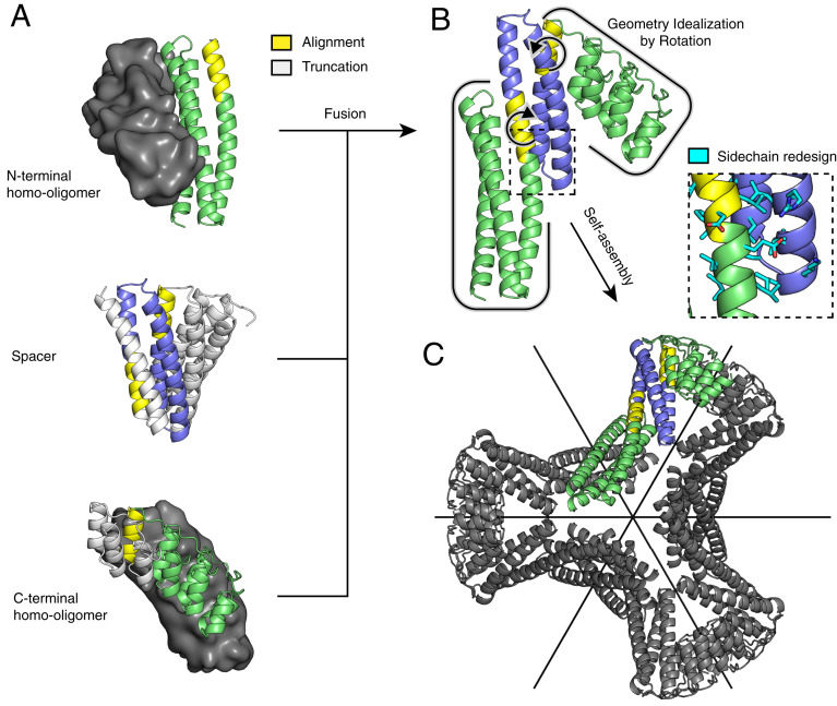
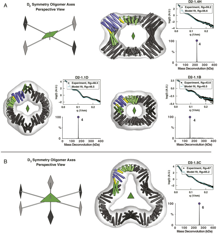
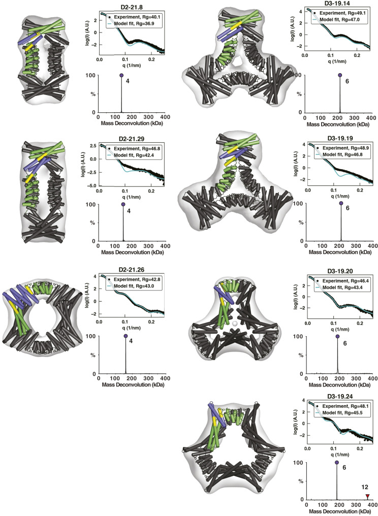
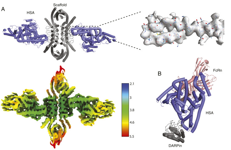
Protein nanomaterial design is an emerging discipline with applications in medicine and beyond. A long-standing design approach uses genetic fusion to join protein homo-oligomer subunits via α-helical linkers to form more complex symmetric assemblies, but this method is hampered by linker flexibility and a dearth of geometric solutions. Here, we describe a general computational method for rigidly fusing homo-oligomer and spacer building blocks to generate user-defined architectures that generates far more geometric solutions than previous approaches. The fusion junctions are then optimized using Rosetta to minimize flexibility. We apply this method to design and test 92 dihedral symmetric protein assemblies using a set of designed homodimers and repeat protein building blocks. Experimental validation by native mass spectrometry, small-angle X-ray scattering, and negative-stain single-particle electron microscopy confirms the assembly states for 11 designs. Most of these assemblies are constructed from designed ankyrin repeat proteins (DARPins), held in place on one end by α-helical fusion and on the other by a designed homodimer interface, and we explored their use for cryogenic electron microscopy (cryo-EM) structure determination by incorporating DARPin variants selected to bind targets of interest. Although the target resolution was limited by preferred orientation effects and small scaffold size, we found that the dual anchoring strategy reduced the flexibility of the target-DARPIN complex with respect to the overall assembly, suggesting that multipoint anchoring of binding domains could contribute to cryo-EM structure determination of small proteins.

Keywords: DARPin; cryo-EM; nanomaterials; protein design; protein fusion.


Conflict of interest statement

The authors declare no competing interest.

Figures

Fig. 1.
Fig. 1.
Illustration of the tripartite design strategy for the D3 architecture. (A) The final structure is composed of two homo-oligomers (dimers, top and bottom; the partner subunit is shown as a surface in gray) and a DHR protein (middle). (B) All possible nonclashing backbone alignments are geometrically analyzed and filtered to generate a three-component fusion, which is idealized to the target geometry by small rigid body rotations and redesigned to improve core packing and remove exposed hydrophobics. (C) The result is a D3 assembly with symmetric C2 axes (black) that correspond to those of the original homodimers and a new C3 axis orthogonal and through the center.
Fig. 2.
Fig. 2.
Characterization of first-round designs. EM, native-MS, and SAXS experiments are consistent with the formation of intended architectures for four designs: three D2 designs (A) and one D3 design (B). Negative-stain 3D reconstructions are overlaid with the design models, whose asymmetric units are colored according to the constituent building blocks (N- and C-terminal oligomers, green; DHR, blue; shared alignment, yellow). Native-MS deconvolutions show the relative abundance of the determined masses, and the peaks are labeled with their assigned oligomeric states. SAXS plots compare the theoretical (cyan) and experimental (black) scattering intensities (log scale) as a function of q as well as radius of gyration (Rg).
Fig. 3.
Fig. 3.
Characterization of second-round designs. EM, native-MS, and SAXS experiments are consistent with the formation of intended architectures for six designs. Negative-stain 3D reconstructions are overlaid by design models, whose asymmetric units are colored according to the constituent building blocks (N- and C-terminal oligomers, green; DHR, blue; shared alignment, yellow). Native-MS deconvolutions show the relative abundance of the determined masses, and the peaks are labeled with their assigned oligomeric states. SAXS plots compare the theoretical (cyan) and experimental (black) scattering intensities (log scale) as a function of q as well as Rg. Native-MS for D3-19.24 shows a small amount of 12-mer, likely formed through the association of two designed hexamers.
Fig. 4.
Fig. 4.
Cryo-EM characterization of DARPin-grafted scaffold with GFP. (A) Design model of four GFP (two in front, two behind) bound to the scaffold D2-1.4H.GFP.v1. GFP-binding DARPin “3G124nc” (24) residues are grafted onto the assembly subunit of D2-1.4H while preserving core residues and homo-oligomer interfaces to form the hybrid structure capable of binding GFP. (B) View of the entire assembly colored by local resolution as calculated by CryoSPARC. Resmap calculates the mean resolution at 5.25 Å and median at 5.0 Å. (C) Surface and clipped (through the central chromophore) views of the GFP β-barrel and DARPin region of the scaffold, colored by local resolution. (D) FSC curves calculated by cryoSPARC. Resolution is 4.78 Å based on the 0.143 FSC level.
Fig. 5.
Fig. 5.
Characterization of anti-HSA DARPin assembly in complex with HSA. (A) A cartoon model of scaffold D2-21.8.HSA-C9.v2 and HSA cocomplex docked into a locally refined cryo-EM map obtained from the map below, which is colored by resolution (Å). The density map surrounding the indicated two helices is shown as a transparent gray surface with the model docked. (B) A model of the relative HSA-binding positions of the DARPin and FcRn is built by superposition of this cryo-EM structure (DARPin scaffold + HSA) and an existing crystal structure of HSA with FcRn (Protein Data Bank 4K71). Several hydrophobic DARPin residues involved in binding are shown as sticks.

Similar articles

Cited by

References

    1. Liu Y., Huynh D. T., Yeates T. O., A 3.8 Å resolution cryo-EM structure of a small protein bound to an imaging scaffold. Nat. Commun. 10, 1864 (2019). - PMC - PubMed
    1. Yao Q., Weaver S. J., Mock J. Y., Jensen G. J., Fusion of DARPin to aldolase enables visualization of small protein by cryo-EM. Structure 27, 1148–1155.e3 (2019). - PMC - PubMed
    1. Marcandalli J., et al. ., Induction of potent neutralizing antibody responses by a designed protein nanoparticle vaccine for respiratory syncytial virus. Cell 176, 1420–1431.e17 (2019). - PMC - PubMed
    1. Ueda G., et al. ., Tailored design of protein nanoparticle scaffolds for multivalent presentation of viral glycoprotein antigens. eLife 9, e57659 (2020). - PMC - PubMed
    1. Padilla J. E., Colovos C., Yeates T. O., Nanohedra: Using symmetry to design self-assembling protein cages, layers, crystals, and filaments. Proc. Natl. Acad. Sci. U.S.A. 98, 2217–2221 (2001). - PMC - PubMed

Publication types

LinkOut - more resources