. 2021 Jun 8;118(23):e2019740118.
doi: 10.1073/pnas.2019740118.

Mitochondrial apoptotic priming is a key determinant of cell fate upon p53 restoration

Affiliations

Mitochondrial apoptotic priming is a key determinant of cell fate upon p53 restoration

Francisco J Sánchez-Rivera et al. Proc Natl Acad Sci U S A. .

Abstract

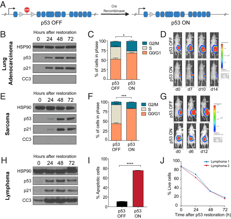
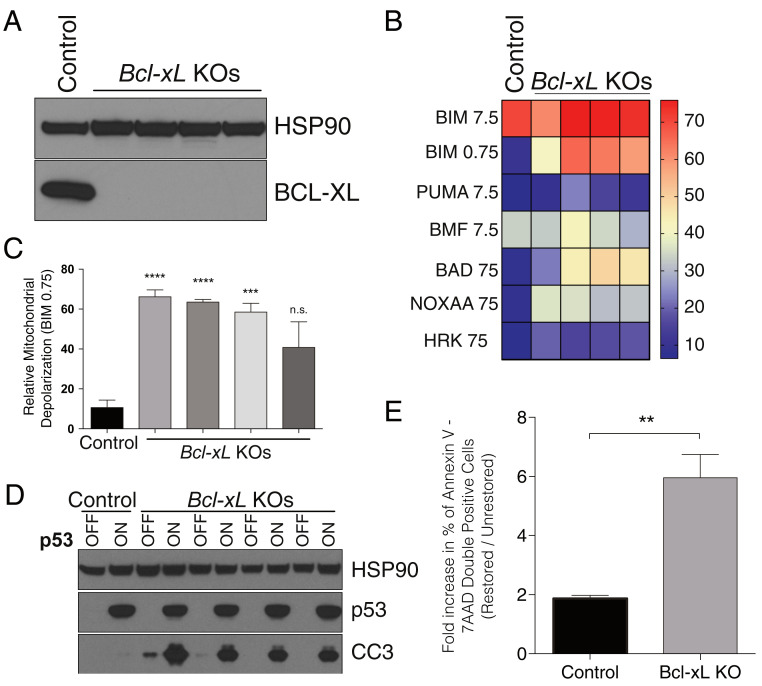
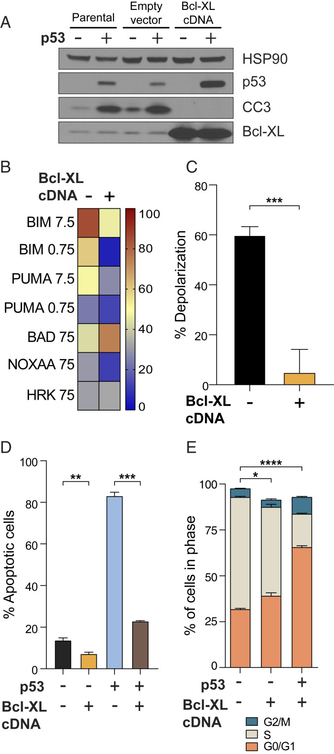
Reactivation of p53 in established tumors typically results in one of two cell fates, cell cycle arrest or apoptosis, but it remains unclear how this cell fate is determined. We hypothesized that high mitochondrial priming prior to p53 reactivation would lead to apoptosis, while low priming would lead to survival and cell cycle arrest. Using a panel of Kras-driven, p53 restorable cell lines derived from genetically engineered mouse models of lung adenocarcinoma and sarcoma (both of which undergo cell cycle arrest upon p53 restoration), as well as lymphoma (which instead undergo apoptosis), we show that the level of mitochondrial apoptotic priming is a critical determinant of p53 reactivation outcome. Cells with high initial priming (e.g., lymphomas) lacked sufficient reserve antiapoptotic capacity and underwent apoptosis after p53 restoration. Forced BCL-2 or BCL-XL expression reduced priming and resulted in survival and cell cycle arrest. Cells with low initial priming (e.g., lung adenocarcinoma and sarcoma) survived and proceeded to arrest in the cell cycle. When primed by inhibition of their antiapoptotic proteins using genetic (BCL-2 or BCL-XL deletion or BAD overexpression) or pharmacologic (navitoclax) means, apoptosis resulted upon p53 restoration in vitro and in vivo. These data demonstrate that mitochondrial apoptotic priming is a key determining factor of cell fate upon p53 activation. Moreover, it is possible to enforce apoptotic cell fate following p53 activation in less primed cells using p53-independent drugs that increase apoptotic priming, including BH3 mimetic drugs.

Keywords: apoptosis; cell cycle arrest; cell fate; p53.


Conflict of interest statement

Competing interest statement: A.L. discloses consulting and sponsored research agreements with AbbVie, Novartis, and Astra-Zeneca. He serves on the Scientific Advisory Board of Flash Therapeutics, Dialectic Therapeutics, and Zentalis Pharmaceuticals. The following are US Patents regarding BH3 profiling owned by Dana-Farber: 10,393,733; 9,902,759; 9,856,303; 9,540,674; 8,221,966; and 7,868,133. A.L. and J.R. are inventors on patent applications US20180128813A1 and US20180120297A1 held/submitted by the Dana-Farber Cancer Institute that covers high-throughput BH3 profiling. T.J. is a member of the Board of Directors of Amgen and Thermo Fisher Scientific. He is also a cofounder of Dragonfly Therapeutics and T2 Biosystems. T.J. serves on the Scientific Advisory Board of Dragonfly Therapeutics, SQZ Biotech, and Skyhawk Therapeutics. None of these affiliations represent a conflict of interest with respect to the design or execution of this study or interpretation of data presented in this report. T.J.’s laboratory currently also receives funding from the Johnson & Johnson Lung Cancer Initiative and The Lustgarten Foundation for Pancreatic Cancer Research, but this funding did not support the research described in this report.

Figures

Fig. 1.
Fig. 1.
Differences in cell fate upon p53 restoration. (A) Schematic of p53 restorable allele. A stop cassette flanked by loxP sites prevents expression of p53 until Cre excises the cassette and restores wild-type p53. (B, E, and H) Western blots showing restoration of p53 and p21 induction in all tumor types but cleavage of caspase 3 only in lymphoma. Three independent samples were tested for each tumor type. (C and F) Cell cycle analysis showing reduction or loss of S-phase cells after p53 restoration in lung adenocarcinoma and sarcoma cells. (D and G) p53 restoration in lung adenocarcinoma and sarcoma tumors in vivo results in growth arrest but not clearance. (I) Loss of viability in lymphoma cells after p53 restoration at one timepoint and (J) over time. *P < 0.05, ***P < 0.001, ****P < 0.0001.
Fig. 2.
Fig. 2.
BH3 profiling reveals differential priming at baseline and during p53 reactivation. (A) Baseline mitochondrial priming profiles of sarcoma, lung adenocarcinoma, and lymphoma cell lines showing increased priming in lymphoma relative to all other cell lines. Three independent samples were tested for each tumor type. (B) Comparison of priming measured by BIM peptide at 0.75 μM showing the difference between lymphoma priming and all other cell lines. ****P < 0.0001; n.s., not significant. (C and D) BH3 profiling showing increase in priming with time after p53 restoration in lung adenocarcinoma and sarcoma cell lines, respectively.
Fig. 3.
Fig. 3.
Genetic priming of lung adenocarcinoma cell lines is sufficient to switch cell fate of p53-restored cells from cell cycle arrest to apoptosis. (A) CRISPR-mediated deletion of Bcl-xL in lung adenocarcinoma cell lines. (B) BH3 profile of four Bcl-xL knockout lung adenocarcinoma cell lines showing that Bcl-xL deletion increases mitochondrial apoptotic priming. (C) Priming measured by BIM peptide is significantly increased in four Bcl-xL knockout lung adenocarcinoma cell lines. (D) Bcl-xL deletion is sufficient to switch cell fate of p53-restored lung adenocarcinoma cells from cell cycle arrest to apoptosis. Data shown is from cells harvested 72 h after p53 restoration. (E) Fold-change in the percentage of Annexin V–7-AAD double-positive control or Bcl-xL knockout cells 72 h after p53 restoration. Data in C and E represent the mean ± SEM, n = 3 or more. Statistics were calculated with two-sided Student’s t test: **P < 0.01, ***P < 0.001, ****P < 0.0001; n.s., not significant.
Fig. 4.
Fig. 4.
Pharmacological priming of lung adenocarcinoma and sarcoma cell lines is sufficient to switch cell fate of p53-restored cells from cell cycle arrest to apoptosis. (A) BH3 profile of lung adenocarcinoma cells after navitoclax addition showing increased priming. (B) Priming measured by BIM peptide is significantly increased upon ABT263 treatment. (C) Lung adenocarcinoma cells show increased cell death as indicated by nuclear degradation. (D) BH3 profile of sarcoma cells after navitoclax addition showing increased priming. (E) Priming measured by BIM peptide is significantly increased upon ABT263 treatment. (F) Sarcoma cells show increased cell death as indicated by nuclear degradation. *P < 0.05, **P < 0.01, *** P < 0.001; ns, not significant.
Fig. 5.
Fig. 5.
Increased mitochondrial priming by forced expression of BAD is sufficient to switch the fate of sarcoma tumors to apoptosis in vivo. (A) Western blot to verify Bad overexpression in stably transduced sarcoma cells. MIG-Empty = empty vector. MIG-Bad = Bad cDNA expressing vector. (B) BH3 profile of sarcoma cells transduced with empty or BAD-expressing vector showing increased priming by forced expression of BAD. (C) Schematic of experiment for assessing cell fate in vivo. (D) Immunohistochemistry of sarcoma sections stained for cleaved caspase 3 to indicate apoptosis. BAD expressing tumors show much greater apoptosis as indicated by the increased number of cells with cleaved caspase 3 staining. (Magnification: D, 4×; D, Inset, 40×.) *P < 0.05, **P < 0.01; ns, not significant.
Fig. 6.
Fig. 6.
Decreased mitochondrial priming by forced expression of BCL-XL is sufficient to switch the fate of p53-restored lymphoma cell lines to cell cycle arrest. (A) Forced expression of BCL-XL prevents caspase 3 cleavage after p53 restoration and (B) globally reduces mitochondrial priming in lymphoma cell lines. (C) Reduction of mitochondrial priming by BCL-XL expression as measured with BIM 0.75 μM. (D and E) Reduction of mitochondrial priming by BCL-XL expression significantly blunts p53 restoration-mediated apoptosis (D) and leads to a statistically significant decrease in the accumulation of S-phase p53-restored lymphoma cells and a concomitant accumulation in G1 and G2 (E). *P < 0.05, **P < 0.01, ***P < 0.001, ****P < 0.0001.
Fig. 7.
Fig. 7.
The fate of p53 restored cells depends on mitochondrial apoptotic priming. (A) Model depicting a scenario in which increasing mitochondrial apoptotic priming decreases the proportion of cells that undergo cell cycle arrest, whereas decreasing priming would increase this proportion. Diagram created with BioRender.com. (B) Pharmacological priming of cancer cells is sufficient to switch cell fate upon p53 restoration: I, Poorly primed sarcoma and lung adenocarcinoma cells increase mitochondrial priming in response to p53 restoration but do not reach the threshold of apoptosis; II, dual inhibition of BCL-XL and BCL-2 by navitoclax increases mitochondrial priming such that additional priming by p53 restoration crosses the apoptotic threshold; III, lymphoma cells have higher basal mitochondrial priming, and additional p53 induced priming is sufficient to cross the apoptotic threshold; IV, reduction of basal mitochondrial priming by expression of an antiapoptotic protein is sufficient to block p53-restored cells from crossing the apoptotic threshold and instead leads to cell cycle arrest and senescence.

References

    1. Polager S., Ginsberg D., p53 and E2f: Partners in life and death. Nat. Rev. Cancer 9, 738–748 (2009). - PubMed
    1. Momand J., Jung D., Wilczynski S., Niland J., The MDM2 gene amplification database. Nucleic Acids Res. 26, 3453–3459 (1998). - PMC - PubMed
    1. Lane D., Levine A., p53 Research: The past thirty years and the next thirty years. Cold Spring Harb. Perspect. Biol. 2, a000893 (2010). - PMC - PubMed
    1. Lu X., Tied up in loops: Positive and negative autoregulation of p53. Cold Spring Harb. Perspect. Biol. 2, a000984 (2010). - PMC - PubMed
    1. Kastenhuber E. R., Lowe S. W., Putting p53 in context. Cell 170, 1062–1078 (2017). - PMC - PubMed

Publication types

MeSH terms

Substances