Observational Study
. 2021 Jun 1;12(1):3270.
doi: 10.1038/s41467-021-23468-3.

Association between confirmed congenital Zika infection at birth and outcomes up to 3 years of life

Affiliations
Observational Study

Association between confirmed congenital Zika infection at birth and outcomes up to 3 years of life

Najeh Hcini et al. Nat Commun. .

Abstract

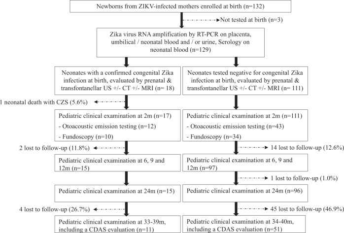
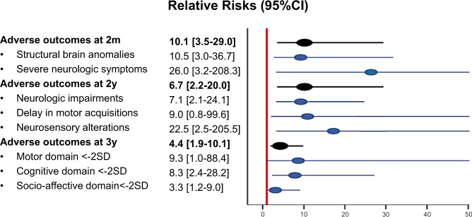
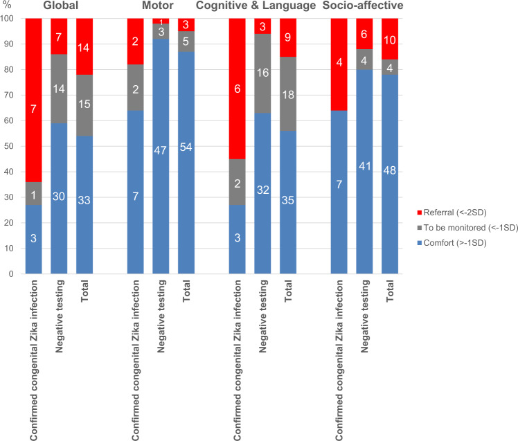
Little is known about the long-term neurological development of children diagnosed with congenital Zika infection at birth. Here, we report the imaging and clinical outcomes up to three years of life of a cohort of 129 children exposed to Zika virus in utero. Eighteen of them (14%) had a laboratory confirmed congenital Zika infection at birth. Infected neonates have a higher risk of adverse neonatal and early infantile outcomes (death, structural brain anomalies or neurologic symptoms) than those who tested negative: 8/18 (44%) vs 4/111 (4%), aRR 10.1 [3.5-29.0]. Neurological impairment, neurosensory alterations or delays in motor acquisition are more common in infants with a congenital Zika infection at birth: 6/15 (40%) vs 5/96 (5%), aRR 6.7 [2.2-20.0]. Finally, infected children also have an increased risk of subspecialty referral for suspected neurodevelopmental delay by three years of life: 7/11 (64%) vs 7/51 (14%), aRR 4.4 [1.9-10.1]. Infected infants without structural brain anomalies also appear to have an increased risk, although to a lesser extent, of neurological abnormalities. It seems paramount to offer systematic testing for congenital ZIKV infection in cases of in utero exposure and adapt counseling based on these results.


Conflict of interest statement

The authors declare no competing interests.

Figures

Fig. 1
Fig. 1. Flowchart of newborns exposed to Zika virus in utero.
All newborns from Zika-infected mothers, living in western French Guiana and followed at the pediatric clinic of the CHOG, were enrolled in this cohort following an informed consent process. At birth, they underwent clinical examination (including anthropometric measurements and a special focus on the neurological status), transfontanellar ultrasound (US), and testing for congenital Zika infection (PCR on urine, blood, and placenta; serology; and testing in cerebrospinal fluid if symptomatic). After postpartum discharge, they were recalled at 2, 6, 9, 12, 18, and 24 months of life for a pediatric examination. At 3 years of life (33–42 months), they were recalled for an evaluation of their development using the Child Development Assessment Scale (CDAS).
Fig. 2
Fig. 2. Main outcomes at 2 months, 2 years, and 3 years of life.
Relative risks of adverse outcomes at 2 months (m), 2 years (y), and 3 years of life, associated with laboratory-confirmed congenital Zika infection at birth are estimated using generalized linear models, adjusted on maternal infection diagnosed in the first trimester, and presented with 95% confidence intervals (95% CI). SD standard deviations.
Fig. 3
Fig. 3. Head circumference and weight in children tested positive and negative for congenital Zika infection at birth.
Head circumferences (in centimeters) and weights (in kilograms) are presented for each time point of this study (birth, 1 month, 2 months, 1 year, 2 years, 3 years). Boxes represent median and interquartile range (IQR), whiskers represent range excluding outliers >1.5× IQR from upper or lower quartile, and circles represent outliers. Z-(blue): Children tested negative at birth for congenital Zika infection; Z+ (red): children tested positive at birth for congenital Zika infection. n = 129 neonates at birth (18 Z+/ 111 Z−), n = 128 infants at 1 and 2 months (17 Z+/ 111 Z−), n = 112 infants at 12 months (15 Z+/ 97 Z−), n = 111 children at 24 months (15 Z+/ 96 Z−), n = 62 children at 36 months (11 Z+/51 Z−).
Fig. 4
Fig. 4. Childhood development at 3 years of life.
All children followed for in utero ZIKV exposure were recalled for a developmental evaluation using the Child Development Assessment Scale at 3 years of life (33–42 months, n = 62). Normal results are classified in the “comfort” or “blue” zone (>−1 standard deviation [SD]). Intermediate results are classified in the “to be monitored” or “gray” zone (−2SD; −1SD). Suspicion of delays is classified in the “referral” or “red” zone (<−2SD). The motor, socio-emotional, and cognitive & language domains were evaluated using this scale. Results of these domains were synthesized in a global evaluation.
Fig. 5
Fig. 5. Healthcare services in western French Guiana.
All newborns from Zika-infected mothers, living in western French Guiana (within the red lines) and followed at the pediatric clinic of the CHOG (large red dot), were enrolled in this cohort. Asymptomatic neonates from mothers living outside of the Saint-Laurent du Maroni area (blue area), were discharged after day 3–5 postpartum and were followed in the nearest primary healthcare center (small red dot).

References

    1. Haddow AD, et al. Genetic characterization of Zika virus strains: geographic expansion of the Asian lineage. PLoS Negl. Trop. Dis. 2012;6:e1477. doi: 10.1371/journal.pntd.0001477. - DOI - PMC - PubMed
    1. Brasil, P., et al. Zika virus outbreak in Rio de Janeiro, Brazil: clinical characterization, epidemiological and virological aspects. PloS Negl. Trop. Dis.10 (2016). - PMC - PubMed
    1. Brasil P, et al. Zika virus infection in pregnant women in Rio de Janeiro. N. Engl. J. Med. 2016;375:2321–2334. doi: 10.1056/NEJMoa1602412. - DOI - PMC - PubMed
    1. Pomar L, et al. Association between Zika virus and fetopathy: a prospective cohort study in French Guiana. Ultrasound Obstet. Gynecol. 2017;49:729–736. doi: 10.1002/uog.17404. - DOI - PubMed
    1. Moore CA, et al. Characterizing the pattern of anomalies in congenital Zika syndrome for pediatric clinicians. JAMA Pediatr. 2017;171:288–295. doi: 10.1001/jamapediatrics.2016.3982. - DOI - PMC - PubMed

Publication types

MeSH terms