. 2021 Jun 1;7(1):20.
doi: 10.1038/s41526-021-00148-6.

3D cell culture using a clinostat reproduces microgravity-induced skin changes

Affiliations

3D cell culture using a clinostat reproduces microgravity-induced skin changes

Dong Hyun Choi et al. NPJ Microgravity. .

Abstract

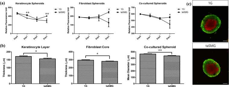
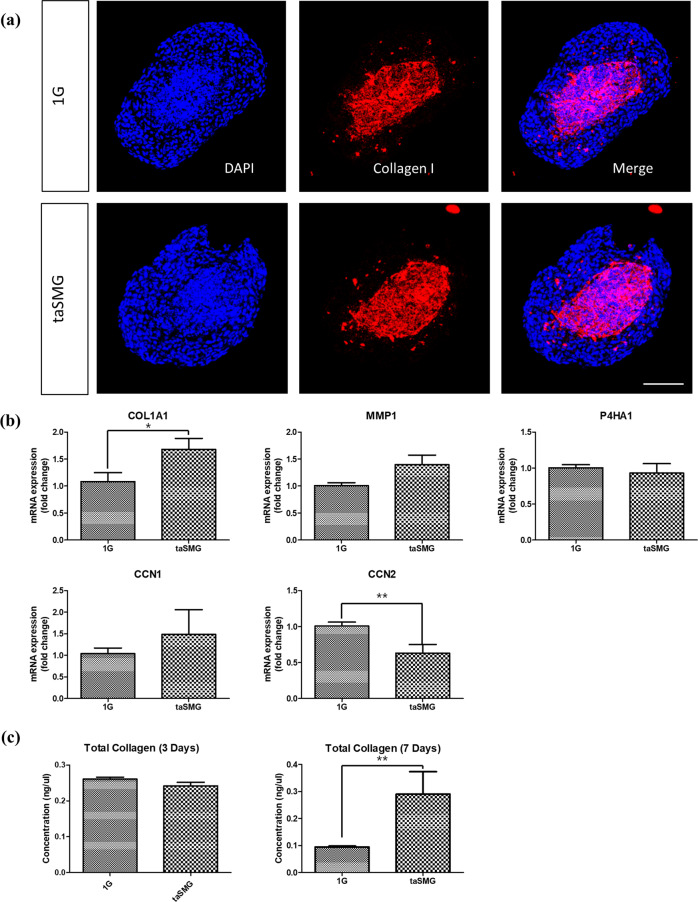
Exposure to microgravity affects human physiology in various ways, and astronauts frequently report skin-related problems. Skin rash and irritation are frequent complaints during space missions, and skin thinning has also been reported after returning to Earth. However, spaceflight missions for studying the physiological changes in microgravity are impractical. Thus, we used a previously developed 3D clinostat to simulate a microgravity environment and investigate whether physiological changes of the skin can be reproduced in a 3D in vitro setting. Our results showed that under time-averaged simulated microgravity (taSMG), the thickness of the endothelial cell arrangement increased by up to 59.75%, indicating skin irritation due to vasodilation, and that the diameter of keratinocytes and fibroblast co-cultured spheroids decreased by 6.66%, representing skin thinning. The α1 chain of type I collagen was upregulated, while the connective tissue growth factor was downregulated under taSMG. Cytokeratin-10 expression was significantly increased in the taSMG environment. The clinostat-based 3D culture system can reproduce physiological changes in the skin similar to those under microgravity, providing insight for understanding the effects of microgravity on human health before space exploration.


Conflict of interest statement

The authors declare no competing interests.

Figures

Fig. 1
Fig. 1. Arrangement of HUVECs under various gravity environments using Matrigel.
After 18 h of incubation under a 1 G, b 1G-to-taSMG, and c taSMG. Scale bars: 100 μm. d Quantitative comparison of thickness between groups. Data represent the mean ± SEM (n = 8–9, *p < 0.05).
Fig. 2
Fig. 2. Cell viability and thickness of spheroids under 1 G and taSMG.
a Cell viability of mono- and co-cultured spheroids for 3 days of culture. Keratinocyte spheroids showed significant difference at day 1 and 3. All spheroids showed a significant difference after 3 days. b Thickness between keratinocyte layer and fibroblast core and overall diameter of co-cultured spheroids. All comparisons between 1 G and taSMG showed significant differences after 3 days. Data represent the mean ± SEM (n = 8, *p < 0.05, **p < 0.01). c Confocal images of CellTrackerTM stained spheroids under 1 G and taSMG. Red tracker indicates fibroblast core and green tracker indicates keratinocyte layer. Scale bars: 100 μm.
Fig. 3
Fig. 3. Extracellular matrix homeostasis of co-cultured spheroids under 1 G and taSMG.
a Collagen type I and DAPI staining showing collagen expression in the fibroblast core and both nuclei of the keratinocyte layer and fibroblast core. Scale bar: 100 μm. b qRT-PCR analysis of COL1A1, MMP-1, P4ha1, CCN1, and CCN2. c Hydroxyproline assay result indicating collagen contents in spheroids after 3 and 7 days. Data represent the mean ± SEM (n = 4, *p < 0.05, **p < 0.01).
Fig. 4
Fig. 4. Cytokeratin-10 (K10) expression of co-cultured spheroids under 1 G and taSMG.
a K10 and DAPI staining showing the cytokeratin expression in keratinocyte layer and both nuclei of the keratinocyte layer and fibroblast core. Scale bar: 100 μm. b qRT-PCR analysis of K10. Data represent the mean ± SEM (n = 4, *p < 0.05).

References

    1. Bloomfield SA, Martinez DA, Boudreaux RD, Mantri AV. Microgravity stress: bone and connective tissue. Compr. Physiol. 2016;6:645–686. doi: 10.1002/cphy.c130027. - DOI - PubMed
    1. Roberts DR, et al. Prolonged microgravity affects human brain structure and function. AJNR Am. J. Neuroradiol. 2019;40:1878–1885. - PMC - PubMed
    1. Shen M, Frishman WH. Effects of spaceflight on cardiovascular physiology and health. Cardiol. Rev. 2019;27:122–126. doi: 10.1097/CRD.0000000000000236. - DOI - PubMed
    1. Crucian B, et al. Incidence of clinical symptoms during long-duration orbital spaceflight. Int. J. Gen. Med. 2016;9:383–391. doi: 10.2147/IJGM.S114188. - DOI - PMC - PubMed
    1. Antonsen, E. et al. Evidence report: risk of adverse health outcomes and decrements in performance due to in-flight medical conditions. National Aeronautics and Space Administration, Houston, TX, USA. Approved for public release: May8 (2017).