. 2021 Jun 1;11(1):11474.
doi: 10.1038/s41598-021-90822-2.

p62 overexpression induces TDP-43 cytoplasmic mislocalisation, aggregation and cleavage and neuronal death

Affiliations

p62 overexpression induces TDP-43 cytoplasmic mislocalisation, aggregation and cleavage and neuronal death

A D Foster et al. Sci Rep. .

Abstract

Amyotrophic lateral sclerosis (ALS) and frontotemporal lobar degeneration (FTLD) that exist on a spectrum of neurodegenerative disease. A hallmark of pathology is cytoplasmic TDP-43 aggregates within neurons, observed in 97% of ALS cases and ~ 50% of FTLD cases. This mislocalisation from the nucleus into the cytoplasm and TDP-43 cleavage are associated with pathology, however, the drivers of these changes are unknown. p62 is invariably also present within these aggregates. We show that p62 overexpression causes TDP-43 mislocalisation into cytoplasmic aggregates, and aberrant TDP-43 cleavage that was dependent on both the PB1 and ubiquitin-associated (UBA) domains of p62. We further show that p62 overexpression induces neuron death. We found that stressors (proteasome inhibition and arsenic) increased p62 expression and that this shifted the nuclear:cytoplasmic TDP-43 ratio. Overall, our study suggests that environmental factors that increase p62 may thereby contribute to TDP-43 pathology in ALS and FTLD.


Conflict of interest statement

The authors declare no competing interests.

Figures

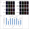
Figure 1
Figure 1
Expression of EGFP-p62 causes TDP-43WT cytoplasmic mislocalisation in a UBA domain dependent manner. Cells co-expressing TDP-43WT and either EGFP or EGFP-p62 (WT or ΔUBA) were fixed and stained with Hoechst then visualized by confocal microscopy. Arrows show TDP-43 forming a shell around wild type p62 (inset). Three independent experiments were performed. Cells expressing both TDP-43-tdTomato and EGFP construct were imaged from random fields of view with a minimum of 10 cells imaged per experiment. TDP-43-tdTomato co-localisation with the nucleus was determined by calculating the Pearson’s co-efficient with ImageJ. Post-hoc ANOVA Bonferroni test was performed in SPSS.
Figure 2
Figure 2
p62 expression alters TDP-43 solubility. Cells were transfected with TDP-43WT and either EGFP, EGFP-p62WT or EGFP-p62ΔUBA (as indicated), and 48 h post-transfection cells were lysed in Triton-X buffer (soluble). Triton-X insoluble pellets were solubilized with SDS (insoluble). Western blot analyses with GFP, RFP and α-tubulin antibodies were performed. Image is representative of 3 independent experiments.
Figure 3
Figure 3
p62 alters the nuclear:cytoplasmic TDP-43 ratio. Mouse embryonic fibroblasts (A) with p62 knockdown (p62−/−) and (B) without (p62+/+) were treated with proteasome inhibitor (MG132), sodium arsenate or heat shock followed by recovery. Cells were fractionated into nuclear and cytosol fractions and proteins separated by SDS polyacrylamide gel electrophoresis. Western blots for endogenous p62, TDP-43 and α-tubulin were performed. (C) The ratio of nuclear: cytoplasmic TDP-43 in non-treated p62−/− cells presented as a fold-change normalised to p62+/+ non-treated cells (set to 1.0) and (D) the amount of p62 in p62+/+ with indicated treatments. (E) The ratio of nuclear:cytoplasmic TDP-43 in treated p62+/+ cells presented as a fold-change normalised to p62+/+ non-treated cells (set to 1.0). Images are representative of 4 independent experiments. Data presented in (C), (D) and (E) are the mean +/− SEM of 4 independent experiments.
Figure 4
Figure 4
p62 expression induces TDP-43 cleavage to a 35-kDa fragment. Cells were transfected with constant TDP-43-tdTomato with increasing amounts of EGFP-p62 (0, 0.25, 0.5, 1, 1.5 or 2 μg) and decreasing amounts of EGFP (2, 1.75, 1.5, 1, 0.5 or 0 μg) to maintain consistent DNA amounts across transfection. Cells were then lysed in Triton-X buffer (soluble) and Triton-X insoluble pellets were solubilized with SDS (insoluble). Western blots for GFP, RFP and α-tubulin were performed as indicated. Image is representative of 3 independent experiments.
Figure 5
Figure 5
p62 creates a cleaved TDP-43 species that is also induced by proteasomal inhibition. Cells were transfected with TDP-43-tdTomato and either pcDNA3.1 (A) or EGFP-p62 (B). Cells were treated with proteasome inhibitor MG132, serum starved, serum starved with Bafilomycin A1 or left untreated. Cells were lysed and soluble and insoluble fractions obtained. Western blots were performed as indicated. Red dashed boxes indicate the presence of a *TDP-43-tdTomato cleavage product (TDP-35-tdTomato). The TDP-35 band from pcDNA3.1 transfected MG132-treated cells and EGFP-p62 non-treated cells was excised, trypsin-digested and analysed by mass spectrometry (Fig. 6). However, we did observe that the presumed TDP-35 fragment induced by EGFP-p62WT overexpression was also present in the lysates from EGFP expressing cells that were treated with the MG132. These protein bands were excised for LC–MS/MS analysis and we identified four tryptic peptides to confirm that the band matching 90-kDa TDP-tdTomato was indeed a cleavage product of TDP-43 (Fig. 6).
Figure 6
Figure 6
Identified tryptic peptides from TDP-43. The 90 kDa-tdTomato-TDP43 bands from cells transfected with the empty vector treated with MG132 (A) and overexpressed with p62 (B) were excised and underwent tryptic digestion. Peptide analysis indicated that the fragments consist of the C-terminal aa85-414 of TDP-43.
Figure 7
Figure 7
p62-mediated cleavage of TDP-43 is independent of caspases. NSC-34 cells were pre-treated with 20 μM caspase inhibitor, 5 μM calpain inhibitor or were left untreated for 1 h and then transfected with TDP-43-tdTomato and either EGFP or EGFP-p62. Cells were maintained in media containing the inhibitor for 48 h and then harvested for soluble and insoluble fractions. Western blot analyses for TDP-43 (anti-RFP), EGFP and α-tubulin were performed. TDP-43 bands were quantified using ImageJ and the amount of TDP-35-tdTomato fragment observed in the insoluble fraction of EGFP versus EGFP-p62 expressing cells was compared with SPSS.
Figure 8
Figure 8
Expression of p62 causes neuronal death. Cells were transfected with EGFP empty vector (EGFP-EV) and RFP empty vector (RFP-EV), EGFP-p62 with or without TDP-43-tdTomato (RFP-TDP) or TDP-43-tdTomato with EGFP-EV. At (A) 48, (B) 72 or (C) 96 h post-transfection cells were trypsinised and incubated with Sytox-Red and assessed for cell viability by flow cytometry, (D) percentage viability of cells expressing EGFP-p62 + RFP-EV compared to EGFP-EV + RFP-EV expressing cells at each time point. Data is the mean of 3 independent experiments +/− SEM.
Figure 9
Figure 9
p62-mediated neuronal death is independent of TDP-43 expression. (A) Cells were transfected with a PMO to knock-down TDP-43 (TDP-43 KD) or a standard control (GTC). The following day cells were transfected with EGFP-p62 and 48 h later trypsinised, incubated with Sytox-Red and then assessed for cell viability by flow cytometry. (B) Cells were harvested at 3 and 5 days post transfection with PMOs and western blots performed as indicated.
Figure 10
Figure 10
p62 overexpression leads to loss of TDP-43-mediated RNA regulation. Effects of p62 overexpression on alternative splicing of selected genes, showing RT-PCRs across Stag2 (A), Poldip3 (B) and Madd (C) transcripts following transfection with p62 overexpression vector (p62) or the empty vector control (EV) and incubated for 1, 3 and 5 days. An RT-PCR across Smn transcripts were used as a loading control. A 100 bp ladder was loaded on either side of the gel and a no template control (NTC) was included in the final lane. Full-length (FL) and exon skipped (Δ) transcripts are annotated and the percentage of specific transcripts are detailed below each lane.
Figure 11
Figure 11
TDP-43 aggregation and cleavage require the PB1 and UBA domains, and the nuclear export signal of p62. NSC-34 cells were transfected with TDP-43-tdTomato and EGFP-p62 wild type [WT] or mutants as indicated. (A) Schematic diagram of p62 domain structure and deletion mutants: 1–122 (PB1 domain only), 124–440 (lacks the PB1 domain), 1–385 (lacks the UBA domain), Δ122–386 (PB1 and UBA domains only), Δ303–320 (lacks the nuclear export sequence) and K7A/D69A (self-dimerization mutant). (B) Soluble and insoluble fractions were prepared and separated by SDS-PAGE and transferred to nitrocellulose membranes. Western blot analyses were performed as indicated. Image is representative of 3 independent experiments.
Figure 12
Figure 12
TDP-43 nuclear depletion requires the PB1 and UBA domains of p62, but not the nuclear export signal or self-dimerisation via the PB1 domain. (A) NSC-34 cells were transfected with TDP-43-tdTomato and EGFP empty vector (EGFP-EV) or EGFP-p62 (WT or deletion constructions as indicated). Cells were fixed and stained with the nuclear stain Hoechst. Images are representative of 3 independent experiments. Minimum number of cells analysed per construct = 32. (B) TDP-43-tdTomato co-localisation with the nucleus was determined by calculating the Pearson’s co-efficient with ImageJ. Statistical analyses were performed using SPSS, with Bonferroni and Dunnet T3, p = 0.05. #compared with EGFP-EV, *compared with EGFP-p62 WT.

Similar articles

Cited by

References

    1. Couratier P, Corcia P, Lautrette G, Nicol M, Marin B. ALS and frontotemporal dementia belong to a common disease spectrum. Rev. Neurol. (Paris) 2017;173(5):273–279. doi: 10.1016/j.neurol.2017.04.001. - DOI - PubMed
    1. Smith EF, Shaw PJ, De Vos KJ. The role of mitochondria in amyotrophic lateral sclerosis. Neurosci. Lett. 2017;710:132933. doi: 10.1016/j.neulet.2017.06.052. - DOI - PubMed
    1. Ramesh N, Pandey UB. Autophagy dysregulation in ALS: When protein aggregates get out of hand. Front. Mol. Neurosci. 2017;10:263. doi: 10.3389/fnmol.2017.00263. - DOI - PMC - PubMed
    1. Purice MD, Taylor JP. Linking hnRNP function to ALS and FTD pathology. Front. Neurosci. 2018;12:326. doi: 10.3389/fnins.2018.00326. - DOI - PMC - PubMed
    1. Ji AL, Zhang X, Chen WW, Huang WJ. Genetics insight into the amyotrophic lateral sclerosis/frontotemporal dementia spectrum. J. Med. Genet. 2017;54(3):145–154. doi: 10.1136/jmedgenet-2016-104271. - DOI - PubMed

Publication types