Case Reports
. 2021 Sep;29(9):1359-1368.
doi: 10.1038/s41431-021-00900-2. Epub 2021 Jun 1.

A MT-TL1 variant identified by whole exome sequencing in an individual with intellectual disability, epilepsy, and spastic tetraparesis

Collaborators, Affiliations
Case Reports

A MT-TL1 variant identified by whole exome sequencing in an individual with intellectual disability, epilepsy, and spastic tetraparesis

Elke de Boer et al. Eur J Hum Genet. 2021 Sep.

Erratum in

Abstract

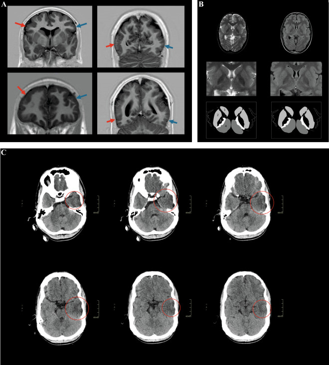
The genetic etiology of intellectual disability remains elusive in almost half of all affected individuals. Within the Solve-RD consortium, systematic re-analysis of whole exome sequencing (WES) data from unresolved cases with (syndromic) intellectual disability (n = 1,472 probands) was performed. This re-analysis included variant calling of mitochondrial DNA (mtDNA) variants, although mtDNA is not specifically targeted in WES. We identified a functionally relevant mtDNA variant in MT-TL1 (NC_012920.1:m.3291T > C; NC_012920.1:n.62T > C), at a heteroplasmy level of 22% in whole blood, in a 23-year-old male with severe intellectual disability, epilepsy, episodic headaches with emesis, spastic tetraparesis, brain abnormalities, and feeding difficulties. Targeted validation in blood and urine supported pathogenicity, with heteroplasmy levels of 23% and 58% in index, and 4% and 17% in mother, respectively. Interestingly, not all phenotypic features observed in the index have been previously linked to this MT-TL1 variant, suggesting either broadening of the m.3291T > C-associated phenotype, or presence of a co-occurring disorder. Hence, our case highlights the importance of underappreciated mtDNA variants identifiable from WES data, especially for cases with atypical mitochondrial phenotypes and their relatives in the maternal line.


Conflict of interest statement

The authors declare no competing interests.

Figures

Fig. 1
Fig. 1. Neuroimaging displays pachygyria, polymicrogyria, white matter abnormalities, and loss of gray–white differentiation.
Coronal MRI images (A) in the phase-sensitive inversion recovery sequence at age 14 years. The images in the upper row show exemplary regions with a microgyrated aspect (red arrow) as compared to the contralateral region (blue arrow). The images in the lower row show exemplary regions with pachygyration (red arrow). In these regions, the gyrus–sulcus pattern is lost as compared to the contralateral side (blue arrow). Polymicrogyria and pachygyria appear most prominent in the frontoparietal cortical areas. Axial T2-weighted MR images (B; left) and fluid attenuation inversion recovery (FLAIR) images (B; right) at the level of basal ganglia at age 14 years. The middle row shows a magnification of the basal ganglia derived from images in the upper row, with a schematic representation in the lower row. 1: caudate nucleus; 2: putamen; 3: globus pallidus; 4: thalamus; dotted line: white matter in between the basal ganglia representing the area of the internal capsule. In the posterior limb of the internal capsule, white hyperintensities are present. These can be recognized by their T2-weighted/FLAIR hyperintense aspect and suggest microstructural white matter degeneration. In addition, the globus pallidus on both sides shows a hypo-intense aspect on the T2-weighted images and FLAIR sequence. Axial CT scan images (C) in brain tissue setting (W/L: 90/40 HU) at age 17 years. Slides should be viewed from left to right to follow the caudocranial axis; the upper-left corner shows the most caudal slide, the lower-right corner shows the most cranial slide. The red circles indicate the hypodense configuration of the temporal lobe with loss of gray–white differentiation. Gray–white differentiation refers to the appearance of the interface between cerebral white matter and cerebral gray matter on brain CT imaging. Loss of gray–white differentiation often indicates the occurrence of cytotoxic edema. In turn, cytotoxic edema is typical for infarction and hypoxic-ischemic encephalopathic syndromes. A clear asymmetry between the left temporal lobe and the right temporal lobe can be observed. The hypodense configuration involves the superior, middle, and inferior temporal gyri (Color figure online).
Fig. 2
Fig. 2. Clinical and radiology images show severe scoliosis, dysmorphisms, and dental crowding.
A Clinical photographs without traction (left) and with traction (middle) showing asymmetry of the chest and rib protrusion on the left side. Radiograph of the vertebral column (right) with anteroposterior view in supine position shows a slight left convex curvature of the upper thoracic spine and severe right convex scoliosis of thoracolumbar spine (Cobb’s angle ±75°) with axial rotation (asymmetric projection of spinous processes and pedicles) and asymmetry of the thoracic cavity. B Frontal and C profile facial photographs of the proband (age 19 years), showing a long face with hypotonic appearance, long palpebral fissures, a prominent nose, and small simple ears. The photograph of the mouth (D) shows crowded teeth with gingival hyperplasia (age 21 years). Facial photographs between age 5 months and age 23 years (E), showing a progression of facial dysmorphisms with advancing age. Only mild dysmorphic features are observed in early childhood, including ptosis and a long philtrum (age 5 months to 4 years). However, the proband develops a progressively pronounced long hypotonic face with open mouth (e.g., photographs at 19 versus 10 versus 2 years of age), and has crowded teeth at age 21 (D), whereas teeth appear less crowded at earlier ages (age 7, 13, and 17 years).

References

    1. Srivastava S, Love-Nichols JA, Dies KA, Ledbetter DH, Martin CL, Chung WK, et al. Meta-analysis and multidisciplinary consensus statement: exome sequencing is a first-tier clinical diagnostic test for individuals with neurodevelopmental disorders. Genet Med. 2019;21:2413–21.. doi: 10.1038/s41436-019-0554-6. - DOI - PMC - PubMed
    1. Zurek B, Ellwanger K, Vissers LELM, Schüle R, Synofzik M, Töpf A, et al. Solve-RD: systematic Pan-European data sharing and collaborative analysis to solve Rare Diseases. Submitted to EJHG (698-20-EJHG). - PMC - PubMed
    1. Matalonga L, Hernández-Ferrer C, Piscia D, Vissers LELM, Schüle R, group S-RS-iw, et al. Solving patients with rare diseases through programmatic reanalysis of genome-phenome data. Submitted to EJHG (703-20-EJHG). - PMC - PubMed
    1. Finsterer J. Genetic, pathogenetic, and phenotypic implications of the mitochondrial A3243G tRNALeu(UUR) mutation. Acta Neurol Scand. 2007;116:1–14. doi: 10.1111/j.1600-0404.2007.00836.x. - DOI - PubMed
    1. Chin J, Marotta R, Chiotis M, Allan EH, Collins SJ. Detection rates and phenotypic spectrum of m.3243A>G in the MT-TL1 gene: a molecular diagnostic laboratory perspective. Mitochondrion. 2014;17:34–41. doi: 10.1016/j.mito.2014.05.005. - DOI - PubMed

Publication types