Review
. 2021 Jul;19(7):409-424.
doi: 10.1038/s41579-021-00573-0. Epub 2021 Jun 1.

SARS-CoV-2 variants, spike mutations and immune escape

Affiliations
Review

SARS-CoV-2 variants, spike mutations and immune escape

William T Harvey et al. Nat Rev Microbiol. 2021 Jul.

Abstract

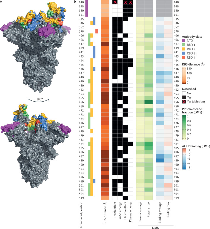
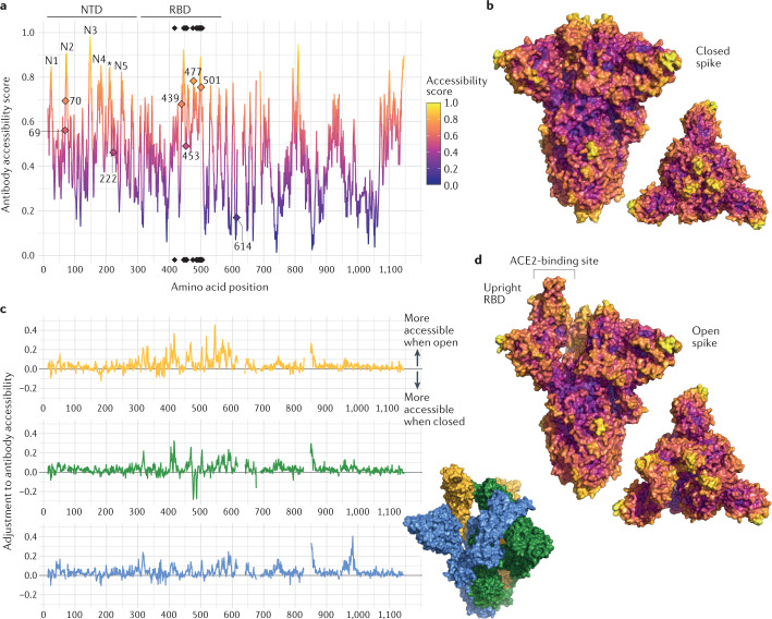
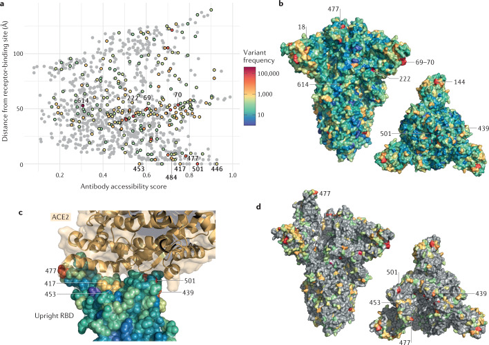
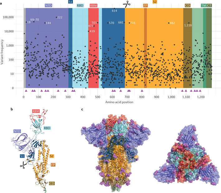
Although most mutations in the severe acute respiratory syndrome coronavirus 2 (SARS-CoV-2) genome are expected to be either deleterious and swiftly purged or relatively neutral, a small proportion will affect functional properties and may alter infectivity, disease severity or interactions with host immunity. The emergence of SARS-CoV-2 in late 2019 was followed by a period of relative evolutionary stasis lasting about 11 months. Since late 2020, however, SARS-CoV-2 evolution has been characterized by the emergence of sets of mutations, in the context of 'variants of concern', that impact virus characteristics, including transmissibility and antigenicity, probably in response to the changing immune profile of the human population. There is emerging evidence of reduced neutralization of some SARS-CoV-2 variants by postvaccination serum; however, a greater understanding of correlates of protection is required to evaluate how this may impact vaccine effectiveness. Nonetheless, manufacturers are preparing platforms for a possible update of vaccine sequences, and it is crucial that surveillance of genetic and antigenic changes in the global virus population is done alongside experiments to elucidate the phenotypic impacts of mutations. In this Review, we summarize the literature on mutations of the SARS-CoV-2 spike protein, the primary antigen, focusing on their impacts on antigenicity and contextualizing them in the protein structure, and discuss them in the context of observed mutation frequencies in global sequence datasets.


Conflict of interest statement

The authors declare no competing interests.

Figures

Fig. 1
Fig. 1. Neutralizing antibody classes defined by structural analyses and properties of spike protein residues.
a | Amino acid residues of the severe acute respiratory syndrome coronavirus 2 (SARS-CoV-2) spike protein are coloured according to the class of the antibody that binds to an epitope. Receptor-binding domain (RBD) antibody classes 1–4 (ref.) are shown: green for class 1 (ACE2-blocking antibodies that bind the spike protein in the open conformation), yellow for class 2 (ACE2-blocking antibodies that bind the RBD in both the open conformation and the closed conformations), blue for class 3 (antibodies that do not block ACE2 and bind the RBD in both the open conformation and the closed conformations) and red for class 4 (neutralizing antibodies that bind outside the ACE2 site and only in the open conformation). When residues belong to epitopes of multiple classes, priority colouring is given to antibodies that block ACE2 and bind the closed spike protein. The amino-terminal domain (NTD) supersite is coloured in magenta. b | Aligned heat maps showing properties of amino acid residues where substitutions affect binding by antibodies in polyclonal human blood plasma or emerge as antibody escape mutations. The distance in angstroms to the ACE2-contacting residues that form the receptor-binding site (RBS) is shown in shades of orange; each residue is classified as having evidence for mutations affecting neutralization by either monoclonal antibodies (mAbs),,, or polyclonal antibodies in plasma from previously infected individuals (convalescent)–,, or vaccinated individuals (‘mAb effect’ and ‘plasma effect’, respectively). A subset of these residues has mutations described as emerging upon exposure (co-incubation) to mAbs,, or plasma, in laboratory experiments (‘mAb emerge’ and ‘plasma emerge’, respectively). When an observation includes a deletion, this is indicated by a red cross. Shades of green depict the results of deep mutational scanning (DMS) experiments where yeast cells expressing RBD mutants are incubated with multiple samples of human convalescent plasma. The escape fraction (that is, a quantitative measure of the extent to which a mutation reduced polyclonal antibody binding) averaged across all amino acid substitutions at a residue (‘plasma average’) and the maximally resistant substitution (‘plasma max’) are indicated. DMS data on ACE2-binding affinity are shown in shades of red or blue representing higher or lower ACE2 affinity, respectively. The mean change in binding affinity averaged across all mutations at each site (‘binding average’) and alternatively the maximally binding mutant (‘binding max’) is shown. Scores represent binding constants (Δlog10 KD) relative to the wild-type reference amino acid.
Fig. 2
Fig. 2. Structure-based analysis of conformational epitopes on the spike protein.
a | Structure-based antibody accessibility scores for each spike protein ectodomain residue in the closed form were calculated with BEpro. Black diamonds at the top and bottom of the plot indicate the positions of ACE2-contacting residues. Accessible amino-terminal domain (NTD) loops N1–N5 are labelled, and a loop falling between these is indicated with an asterisk. b | Two surface colour representations of antibody accessibility scores for the spike protein in the closed conformation according to the colour scheme in part a: a trimer axis vertical view (left) and an orthogonal top-down view along this axis (right). c | The extent to which each spike residue becomes more or less accessible when the spike protein is in its open form is shown. For each spike monomer (upright receptor-binding domain (RBD) (yellow), closed RBD clockwise adjacent (green) and closed RBD anticlockwise adjacent (blue)), the difference relative to the score calculated for the closed form (shown in part a) is shown. d | Two surface colour representations of antibody accessibility scores for the spike protein in the open conformation with a single monomer with an upright RBD are shown: a trimer axis vertical view (left) and an orthogonal top-down view along this axis (right).
Fig. 3
Fig. 3. Structural context of spike amino acid mutations in the global virus population.
Spike amino acid residues are coloured according to the frequency of amino acid substitutions or deletions. Variants (retrieved from CoV-GLUE) are based on 426,623 high-quality sequences downloaded from the Global Initiative on Sharing All Influenza Data (GISAID) database on 3 February 2021. a | Points representing each spike amino acid residue are positioned according to the antibody accessibility score and the distance to the nearest residue in the receptor-binding site. Residues with at least 100 sequences possessing a substitution or deletion are coloured according to the frequency scale shown, with the remainder shaded grey. b | Spike protein in closed form with all residues coloured according to the frequency scale shown; a trimer axis vertical view (left) and an orthogonal top-down view along this axis (right) are shown. c | A close-up view of the receptor-binding domain (RBD) bound to ACE2 (RCSB Protein Data Bank ID 6M0J), with RBD residues shown as spheres coloured by amino acid variant frequency and ACE2 shown in gold. Amino acid variants are present at high frequency in positions at the RBD–ACE2 interface. d | Spike protein in open form with residues where at least 100 sequences possessing a substitution are highlighted; a trimer axis vertical view (left) and an orthogonal top-down view along this axis (right) are shown.
Fig. 4
Fig. 4. Spike protein sequence variability and structure.
a | The domain organization of the severe acute respiratory syndrome coronavirus 2 (SARS-CoV-2) spike protein showing amino acid sequence variability. The spike protein is synthesized as a 1,273 amino acid polypeptide, and the frequency of amino acid variants, including both substitutions and deletions, at each of the positions is shown. These variants, relative to the Wuhan-Hu-1 reference sequence, were identified with use of CoV-GLUE, which filters out Global Initiative on Sharing All Influenza Data (GISAID) sequences identified as being of low quality or from non-human hosts (sequences retrieved from the GISAID database on 3 February 2021). Among 426,623 genomes after filtering, 5,106 different amino acid replacements or substitutions across 1,267 spike positions were identified, of which 320 at 259 positions were observed in at least 100 sequences. In addition to substitutions, several deletions have been observed, particularly within the amino-terminal domain (NTD). The most frequently detected NTD deletion is the two-residue deletion at positions 69 and 70 (Δ69–70), present in 45,898 sequences. The S1–S2 boundary is at amino acid position 685. b | Spike protein monomer displaying an upright receptor-binding domain (RBD). c | Spike protein structure in the closed conformation overlaid with surface representations shown with a trimer axis vertical view (left) and an orthogonal top-down view along this axis (right). Domains are coloured as in part a. The RCSB Protein Data Bank IDs for the SARS-CoV-2 spike protein structures are 6ZGG and 6ZGE. The magenta spheres represent glycans, and the magenta triangles represent potantial N-linked glycosylation sites. The scissors represent the S1–S2 boundary at amino acid position 685. CD, connecting domain; CT cytoplasmic tail; FP, fusion peptide; RBM, receptor-binding motif; TM, transmembrane domain.
Fig. 5
Fig. 5. Amino acid mutations that characterize variants of concern.
a | Spike heterotrimer in the open conformation overlaid with the surface representation (RCSB Protein Data Bank ID 6ZGG). The locations of amino acid substitutions and deletions that define variants of concern are highlighted as red spheres. For B.1.1.7, scissors mark the approximate position of substitution P681H within the furin cleavage site, which is absent from the structural model. b | Aligned heat maps showing properties of amino acid residues or of the specific amino acid substitution, as appropriate. Epitope residues are coloured to indicate the amino-terminal domain (NTD) or the receptor-binding domain (RBD) class. Structure-based antibody access scores for the spike protein in the closed and open conformations are shown. For RBD residues, the results of deep mutational scanning (DMS) studies show the escape fraction (that is, a quantitative measure of the extent to which a mutation reduced polyclonal antibody binding) for each mutant averaged across plasma (‘plasma average’) and for the most sensitive plasma (‘plasma max’). Each mutation is classified as having evidence for mutations affecting neutralization by either monoclonal antibodies (mAbs) or antibodies in convalescent plasma or vaccinated individuals, and emerging in selection experiments using mAbs,, or post-infection serum,,. The distance to the ACE2-contacting residues that form the receptor-binding site RBS is shown (for residue 681, this is estimated with use of the nearest residues present in published structures). DMS data on ACE2-binding affinity are shown by aggregation of scores and averaging across each mutant at a residue and alternatively the maximally binding mutant. Scores represent the binding constant (Δlog10 KD) relative to the wild-type reference amino acid. Mutations that are present in a variant but that are also widespread in the virus population in which a variant emerged, or exhibit high diversity within a lineage, are marked with a dagger.

References

    1. WHO. Coronavirus (COVID-19) Dashboard. https://covid19.who.int/ (2021).
    1. Meredith LW, et al. Rapid implementation of SARS-CoV-2 sequencing to investigate cases of health-care associated COVID-19: a prospective genomic surveillance study. Lancet Infect. Dis. 2020;20:1263–1272. - PMC - PubMed
    1. Rambaut, A. et al. Preliminary Genomic Characterisation of an Emergent SARS-CoV-2 Lineage in the UK Defined by a Novel set of Spike Mutations. https://virological.org/t/preliminary-genomic-characterisation-of-an-eme... (2020).
    1. Frost SDW, Magalis BR, Kosakovsky Pond SL. Neutral theory and rapidly evolving viral pathogens. Mol. Biol. Evol. 2018;35:1348–1354. - PMC - PubMed
    1. MacLean OA, Orton RJ, Singer JB, Robertson DL. No evidence for distinct types in the evolution of SARS-CoV-2. Virus Evol. 2020 doi: 10.1093/ve/veaa034. - DOI - PMC - PubMed

Publication types

MeSH terms