

Molecular Aspects Concerning the Use of the SARS-CoV-2 Receptor Binding Domain as a Target for Preventive Vaccines
- PMID: 34075345
- PMCID: PMC8084267
- DOI: 10.1021/acscentsci.1c00216
Molecular Aspects Concerning the Use of the SARS-CoV-2 Receptor Binding Domain as a Target for Preventive Vaccines
Abstract
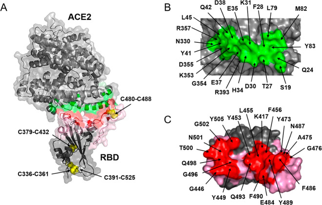
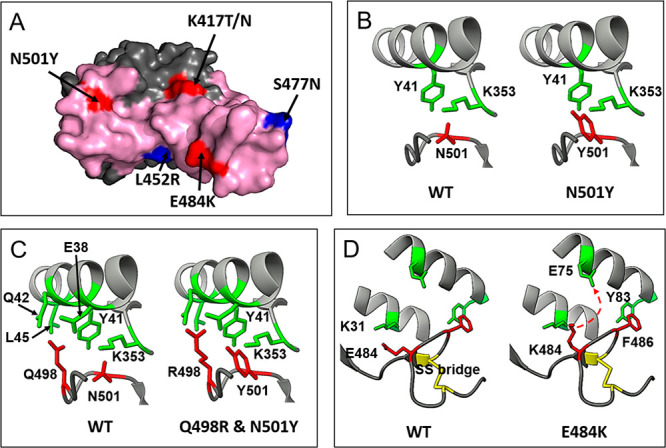
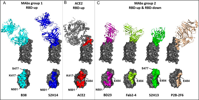
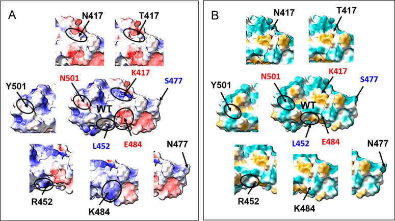
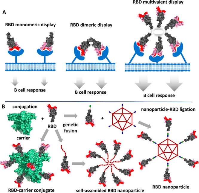
The development of recombinant COVID-19 vaccines has resulted from scientific progress made at an unprecedented speed during 2020. The recombinant spike glycoprotein monomer, its trimer, and its recombinant receptor-binding domain (RBD) induce a potent anti-RBD neutralizing antibody response in animals. In COVID-19 convalescent sera, there is a good correlation between the antibody response and potent neutralization. In this review, we summarize with a critical view the molecular aspects associated with the interaction of SARS-CoV-2 RBD with its receptor in human cells, the angiotensin-converting enzyme 2 (ACE2), the epitopes involved in the neutralizing activity, and the impact of virus mutations thereof. Recent trends in RBD-based vaccines are analyzed, providing detailed insights into the role of antigen display and multivalence in the immune response of vaccines under development.
© 2021 The Authors. Published by American Chemical Society.
Conflict of interest statement
The authors declare the following competing financial interest(s): Y.V.-B., D.S.-M, S.F., L.R., B.S.-R., K.L., D.G.-R., D.G.R., and V.V.B. are co-inventors on provisional SARS-CoV-2 vaccine patents including results covered here.
Figures
Similar articles
-
Structural Basis of a Human Neutralizing Antibody Specific to the SARS-CoV-2 Spike Protein Receptor-Binding Domain.Microbiol Spectr. 2021 Oct 31;9(2):e0135221. doi: 10.1128/Spectrum.01352-21. Epub 2021 Oct 13. Microbiol Spectr. 2021. PMID: 34643438 Free PMC article.
-
A Combination of Receptor-Binding Domain and N-Terminal Domain Neutralizing Antibodies Limits the Generation of SARS-CoV-2 Spike Neutralization-Escape Mutants.mBio. 2021 Oct 26;12(5):e0247321. doi: 10.1128/mBio.02473-21. Epub 2021 Oct 5. mBio. 2021. PMID: 34607456 Free PMC article.
-
Competitive SARS-CoV-2 Serology Reveals Most Antibodies Targeting the Spike Receptor-Binding Domain Compete for ACE2 Binding.mSphere. 2020 Sep 16;5(5):e00802-20. doi: 10.1128/mSphere.00802-20. mSphere. 2020. PMID: 32938700 Free PMC article.
-
Interactions of angiotensin-converting enzyme-2 (ACE2) and SARS-CoV-2 spike receptor-binding domain (RBD): a structural perspective.Mol Biol Rep. 2023 Mar;50(3):2713-2721. doi: 10.1007/s11033-022-08193-4. Epub 2022 Dec 23. Mol Biol Rep. 2023. PMID: 36562937 Free PMC article. Review.
-
Recognition of the SARS-CoV-2 receptor binding domain by neutralizing antibodies.Biochem Biophys Res Commun. 2021 Jan 29;538:192-203. doi: 10.1016/j.bbrc.2020.10.012. Epub 2020 Oct 10. Biochem Biophys Res Commun. 2021. PMID: 33069360 Free PMC article. Review.
Cited by
-
SARS-CoV-2 spike opening dynamics and energetics reveal the individual roles of glycans and their collective impact.Commun Biol. 2022 Nov 3;5(1):1170. doi: 10.1038/s42003-022-04138-6. Commun Biol. 2022. PMID: 36329138 Free PMC article.
-
Chimeric Antigen by the Fusion of SARS-CoV-2 Receptor Binding Domain with the Extracellular Domain of Human CD154: A Promising Improved Vaccine Candidate.Vaccines (Basel). 2022 Jun 3;10(6):897. doi: 10.3390/vaccines10060897. Vaccines (Basel). 2022. PMID: 35746505 Free PMC article.
-
Molecular Level Dissection of Critical Spike Mutations in SARS-CoV-2 Variants of Concern (VOCs): A Simplified Review.ChemistrySelect. 2021 Aug 20;6(31):7981-7998. doi: 10.1002/slct.202102074. Epub 2021 Aug 17. ChemistrySelect. 2021. PMID: 34541298 Free PMC article. Review.
-
Comparative assessment of a COVID-19 vaccine after technology transfer to Iran from critical quality attributes to clinical and immunogenicity aspects.Sci Rep. 2024 Nov 5;14(1):26793. doi: 10.1038/s41598-024-77331-8. Sci Rep. 2024. PMID: 39501012 Free PMC article.
-
A recombinant SARS-CoV-2 receptor-binding domain expressed in an engineered fungal strain of Thermothelomyces heterothallica induces a functional immune response in mice.Vaccine. 2022 Feb 16;40(8):1162-1169. doi: 10.1016/j.vaccine.2022.01.007. Epub 2022 Jan 19. Vaccine. 2022. PMID: 35078661 Free PMC article.
References
-
- Zhou P.; Yang X. L.; Wang X. G.; Hu B.; Zhang L.; Zhang W.; Si H. R.; Zhu Y.; Li B.; Huang C. L.; Chen H. D.; Chen J.; Luo Y.; Guo H.; Jiang R. D.; Liu M. Q.; Chen Y.; Shen X. R.; Wang X.; Zheng X. S.; Zhao K.; Chen Q. J.; Deng F.; Liu L. L.; Yan B.; Zhan F. X.; Wang Y. Y.; Xiao G. F.; Shi Z. L. A pneumonia outbreak associated with a new coronavirus of probable bat origin. Nature 2020, 579 (7798), 270–273. 10.1038/s41586-020-2012-7. - DOI - PMC - PubMed
-
- Muus C.; Luecken M. D.; Eraslan G.; Waghray A.; Heimberg G.; Sikkema L.; Kobayashi Y.; Vaishnav E. D.; Subramanian A.; Smilie C.; Jagadeesh K.; Duong E. T.; Fiskin E.; Triglia E. T.; Ansari M.; Cai P.; Lin B.; Buchanan J.; Chen S.; Shu J.; Haber A. L.; Chung H.; Montoro D. T.; Adams T.; Aliee H.; Samuel J.; Andrusivova A. Z.; Angelidis I.; Ashenberg O.; Bassler K.; Bécavin C.; Benhar I.; Bergenstråhle J.; Bergenstråhle L.; Bolt L.; Braun E.; Bui L. T.; Chaffin M.; Chichelnitskiy E.; Chiou J.; Conlon T. M.; Cuoco M. S.; Deprez M.; Fischer D. S.; Gillich A.; Gould J.; Guo M.; Gutierrez A. J.; Habermann A. C.; Harvey T.; He P.; Hou X.; Hu L.; Jaiswal A.; Jiang P.; Kapellos T.; Kuo C. S.; Larsson L.; Leney-Greene M. A.; Lim K.; Litviňuková M.; Lu J.; Ludwig L. S.; Luo W.; Maatz H.; Madissoon E.; Mamanova L.; Manakongtreecheep K.; Marquette C.-H.; Mbano I.; McAdams A. M.; Metzger R. J.; Nabhan A. N.; Nyquist S. K.; Penland L.; Poirion O. B.; Poli S.; Qi C.; Queen R.; Reichart D.; Rosas I.; Schupp J.; Sinha R.; Sit R. V.; Slowikowski K.; Slyper M.; Smith N.; Sountoulidis A.; Strunz M.; Sun D.; Talavera-López C.; Tan P.; Tantivit J.; Travaglini K. J.; Tucker N. R.; Vernon K.; Wadsworth M. H.; Waldman J.; Wang X.; Yan W.; Zhao W.; Ziegler C. G. K. Integrated analyses of single-cell atlases reveal age, gender, and smoking status associations with cell type-specific expression of mediators of SARS-CoV-2 viral entry and highlights inflammatory programs in putative target cells. bioRxiv 2020, 10.1101/2020.04.19.049254. - DOI
Publication types
LinkOut - more resources
Full Text Sources
Other Literature Sources
Miscellaneous
