. 2021 Sep;30(9):1882-1894.
doi: 10.1002/pro.4139. Epub 2021 Jun 21.

Characterization of an aminotransferase from Acanthamoeba polyphaga Mimivirus

Affiliations

Characterization of an aminotransferase from Acanthamoeba polyphaga Mimivirus

Chase A Seltzner et al. Protein Sci. 2021 Sep.

Abstract

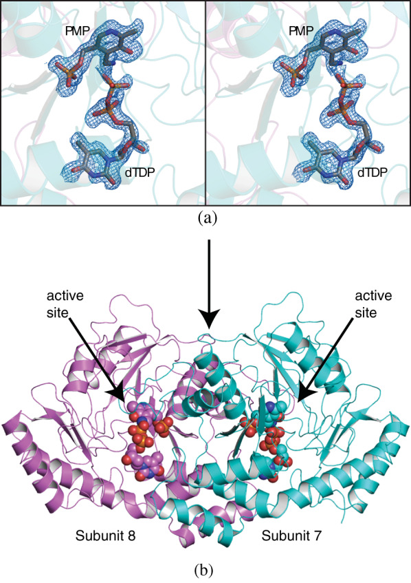
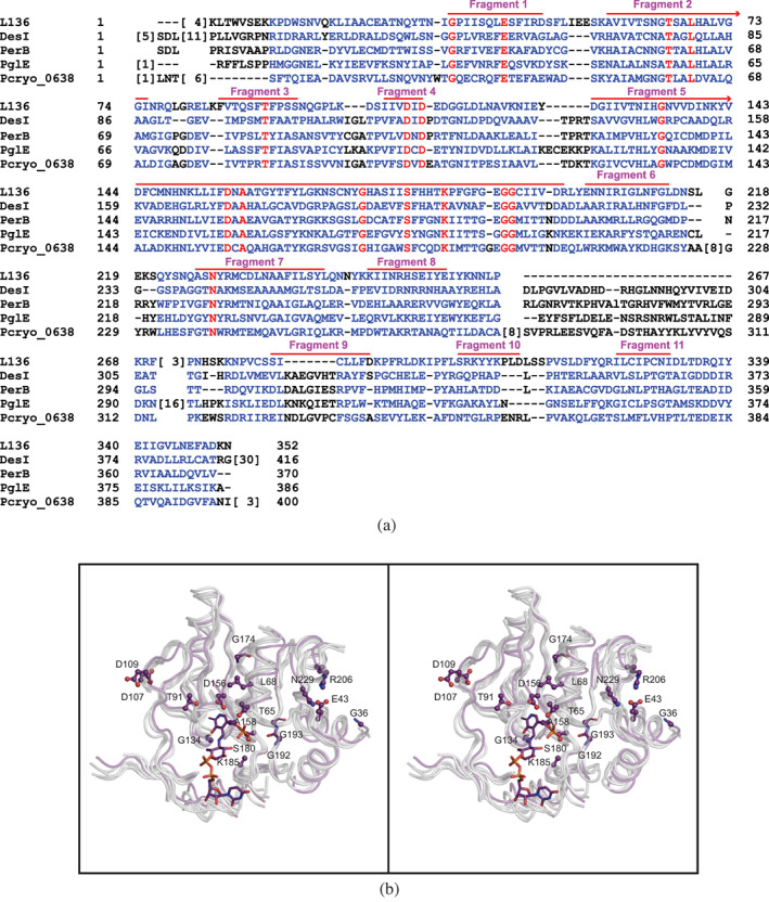
Acanthamoeba polyphaga Mimivirus, a complex virus that infects amoeba, was first reported in 2003. It is now known that its DNA genome encodes for nearly 1,000 proteins including enzymes that are required for the biosynthesis of the unusual sugar 4-amino-4,6-dideoxy-d-glucose, also known as d-viosamine. As observed in some bacteria, the pathway for the production of this sugar initiates with a nucleotide-linked sugar, which in the Mimivirus is thought to be UDP-d-glucose. The enzyme required for the installment of the amino group at the C-4' position of the pyranosyl moiety is encoded in the Mimivirus by the L136 gene. Here, we describe a structural and functional analysis of this pyridoxal 5'-phosphate-dependent enzyme, referred to as L136. For this analysis, three high-resolution X-ray structures were determined: the wildtype enzyme/pyridoxamine 5'-phosphate/dTDP complex and the site-directed mutant variant K185A in the presence of either UDP-4-amino-4,6-dideoxy-d-glucose or dTDP-4-amino-4,6-dideoxy-d-glucose. Additionally, the kinetic parameters of the enzyme utilizing either UDP-d-glucose or dTDP-d-glucose were measured and demonstrated that L136 is efficient with both substrates. This is in sharp contrast to the structurally related DesI from Streptomyces venezuelae, whose three-dimensional architecture was previously reported by this laboratory. As determined in this investigation, DesI shows a profound preference in its catalytic efficiency for the dTDP-linked sugar substrate. This difference can be explained in part by a hydrophobic patch in DesI that is missing in L136. Notably, the structure of L136 reported here represents the first three-dimensional model for a virally encoded PLP-dependent enzyme and thus provides new information on sugar aminotransferases in general.

Keywords: d-viosamine; 4,6-dideoxy sugar; 4-aminoquinovose; Acanthamoeba polyphaga Mimivirus; UDP-4-amino-4,6-dideoxy-d-glucose; X-ray structure; aminotransferase; giant viruses; viral glycans.


Conflict of interest statement

The authors have no competing financial interests.

Figures

SCHEME 1
SCHEME 1
Pathway for the biosynthesis of UDP‐4‐amino‐4,6‐dideoxy‐D‐glucose (UDP‐D‐viosamine)
FIGURE 1
FIGURE 1
Plot of initial velocities versus substrate concentration for L136. Figure 1(a) and (b) are the initial velocity curves utilizing either UDP‐4‐keto‐4,6‐dideoxy‐d‐glucose or dTDP‐4‐keto‐4,6‐dideoxy‐d‐glucose, respectively, as the substrate. Note that measuring velocities over a wide range of substrate concentrations allows us to obtain data that define both k cat and k cat/K M well, which is not accomplished by measuring replicates at fewer different concentrations. The graph shown allows for a qualitative appreciation of the quality of the data; the quantitative goodness‐of‐fit to the Michaelis–Menten equation is given by the standard errors as described in Materials and Methods
FIGURE 2
FIGURE 2
Structure of the wildtype enzyme in complex with PMP and dTDP. Shown in stereo in (a) is the observed electron density corresponding to the bound dTDP and PMP ligands. The map was calculated with (F oF c) coefficients and contoured at 3σ. The ligand was not included in the X‐ray coordinate file used to calculate the omit map, and thus there is no model bias. A ribbon representation of the L136 dimer is presented in (b)
FIGURE 3
FIGURE 3
Structure of the K185A variant with the bound external aldimine formed with UDP‐4‐amino‐4,6‐dideoxy‐d‐glucose. The observed electron density, shown in stereo in (a), was calculated with (F oF c) coefficients and contoured at 3σ. A close‐up view of the active site is displayed in (b). Those side chains displayed in teal are contributed by subunit 1 whereas those colored in pink are provided by subunit 2. Ordered water molecules are displayed as red spheres, and possible hydrogen bonding interactions within 3.2 Å are indicated by the dashed lines
FIGURE 4
FIGURE 4
Structure of the K185A variant with the bound external aldimine formed with dTDP‐4‐amino‐4,6‐dideoxy‐d‐glucose. The observed electron density, shown in stereo in (a), was calculated with (F oF c) coefficients and contoured at 3σ. A superposition of the external aldimines formed with either UDP‐4‐amino‐4,6‐dideoxy‐d‐glucose (in gray) or dTDP‐4‐amino‐4,6‐dideoxy‐d‐glucose (in green) is displayed in (b). The stereo view focuses near the nucleotidyl groups of the external aldimines
SCHEME 2
SCHEME 2
Reaction products for various sugar aminotransferases to be compared
FIGURE 5
FIGURE 5
Plot of initial velocities versus substrate concentration for DesI. Figure 1(a) and (b) are the initial velocity curves utilizing either UDP‐4‐keto‐4,6‐dideoxy‐d‐glucose or dTDP‐4‐keto‐4,6‐dideoxy‐d‐glucose, respectively, as the substrate
FIGURE 6
FIGURE 6
Differences in the binding of the ligands in L136 versus DesI. Shown in stereo is a close‐up view of the protein regions surrounding the nucleotidyl portions of the external aldimines in L136 (white bonds) and DesI (green). Those residue labels with asterisks are contributed by the second subunit of the dimer
FIGURE 7
FIGURE 7
Comparison of L136 with DesI, PerB, PglE, and Pcryo_O638. An amino acid sequence alignment, constructed with the software package COBALT, is provided in (a). There are seven structurally conserved fragment in these enzymes that form the “core” of the aminotransferase structure as presented in stereo in (b). The purple α‐carbon trace corresponds to that of L136 whereas all the other enzymes are highlighted in white. The amino acid side chains shown correspond to those found in L136
FIGURE 8
FIGURE 8
Binding of the external aldimines in L136, DesI, PerB, PglE, and Pcryo_O638. The positions of the external aldimines in L136, DesI, PerB, PglE, and Pcryo_O638 are shown in stereo and are color‐coded in violet, teal, blue, pink, and wheat, respectively

References

    1. Bos L. The embryonic beginning of virology: Unbiased thinking and dogmatic stagnation. Arch Virol. 1995;140:613–619. - PubMed
    1. Shope RE. Thomas Milton RIVERS, 1888‐1962. J Bacteriol. 1962;84:385–388. - PMC - PubMed
    1. La Scola B, Audic S, Robert C, et al. A giant virus in amoebae. Science. 2003;299:2033. - PubMed
    1. Xiao C, Chipman PR, Battisti AJ, et al. Cryo‐electron microscopy of the giant Mimivirus . J Mol Biol. 2005;353:493–496. - PubMed
    1. Raoult D, Audic S, Robert C, et al. The 1.2‐megabase genome sequence of Mimivirus . Science. 2004;306:1344–1350. - PubMed

Publication types

MeSH terms