

Cooperativity of α-Synuclein Binding to Lipid Membranes
- PMID: 34076426
- PMCID: PMC8291482
- DOI: 10.1021/acschemneuro.1c00006
Cooperativity of α-Synuclein Binding to Lipid Membranes
Abstract
Cooperative binding is a key feature of metabolic pathways, signaling, and transport processes. It provides tight regulation over a narrow concentration interval of a ligand, thus enabling switching to be triggered by small concentration variations. The data presented in this work reveal strong positive cooperativity of α-synuclein binding to phospholipid membranes. Fluorescence cross-correlation spectroscopy, confocal microscopy, and cryo-TEM results show that in excess of vesicles α-synuclein does not distribute randomly but binds only to a fraction of all available vesicles. Furthermore, α-synuclein binding to a supported lipid bilayer observed with total internal reflection fluorescence microscopy displays a much steeper dependence of bound protein on total protein concentration than expected for independent binding. The same phenomenon was observed in the case of α-synuclein binding to unilamellar vesicles of sizes in the nm and μm range as well as to flat supported lipid bilayers, ruling out that nonuniform binding of the protein is governed by differences in membrane curvature. Positive cooperativity of α-synuclein binding to lipid membranes means that the affinity of the protein to a membrane is higher where there is already protein bound compared to a bare membrane. The phenomenon described in this work may have implications for α-synuclein function in synaptic transmission and other membrane remodeling events.
Keywords: Adair equation; Cooperative binding; fluorescence correlation spectroscopy; homotropic allostery; lipid membrane; α-synuclein.
Conflict of interest statement
The authors declare no competing financial interest.
Figures

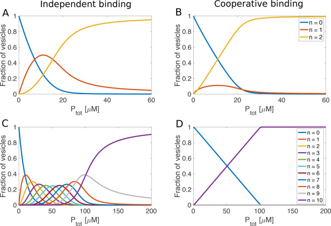
 × 106 M–1 where N is the total number of binding sites. Values
of the macroscopic binding constants for positively cooperative binding
to vesicles with two binding sites were K1 =
× 106 M–1 where N is the total number of binding sites. Values
of the macroscopic binding constants for positively cooperative binding
to vesicles with two binding sites were K1 =  × 106 M–1 and K2 =
4 × 10⁶ M–1, assuming an average
affinity of 1 × 10⁶ M-1 and a free
energy coupling of ΔΔG=-10
kJ mol-1. For the case of vesicles with 10 binding
sites and cooperative binding, the macroscopic binding constants were
K1 = 0.0745 M-1, K2 = 2.15
M-1, K3 = 81.4 M-1,
K4 = 3400 M-1, K5 = 1.5 ×
105 M-1, K6 = 6.7 × 10⁶ M-1, K7 = 2.9 × 10⁸ M-1, K8 = 1.2 × 10¹⁰ M-1, K9 = 4.7 × 10¹¹ M-1 and K10 = 1.3 × 10¹³ M-1, assuming an average affinity of 1
× 106 M-1 and a free energy coupling
of ΔΔG=-10 kJ mol-1.
× 106 M–1 and K2 =
4 × 10⁶ M–1, assuming an average
affinity of 1 × 10⁶ M-1 and a free
energy coupling of ΔΔG=-10
kJ mol-1. For the case of vesicles with 10 binding
sites and cooperative binding, the macroscopic binding constants were
K1 = 0.0745 M-1, K2 = 2.15
M-1, K3 = 81.4 M-1,
K4 = 3400 M-1, K5 = 1.5 ×
105 M-1, K6 = 6.7 × 10⁶ M-1, K7 = 2.9 × 10⁸ M-1, K8 = 1.2 × 10¹⁰ M-1, K9 = 4.7 × 10¹¹ M-1 and K10 = 1.3 × 10¹³ M-1, assuming an average affinity of 1
× 106 M-1 and a free energy coupling
of ΔΔG=-10 kJ mol-1.References
-
- Monod J. (1974) On chance and necessity. In Studies in the Philosophy of Biology, pp 357–375, Springer.
Publication types
MeSH terms
Substances
LinkOut - more resources
Full Text Sources
