Review
. 2021 Jun 24;64(12):7926-7962.
doi: 10.1021/acs.jmedchem.0c02265. Epub 2021 Jun 2.

Recent Advances in the Development of Sigma Receptor Ligands as Cytotoxic Agents: A Medicinal Chemistry Perspective

Affiliations
Review

Recent Advances in the Development of Sigma Receptor Ligands as Cytotoxic Agents: A Medicinal Chemistry Perspective

Antonino N Fallica et al. J Med Chem. .

Abstract

Since their discovery as distinct receptor proteins, the specific physiopathological role of sigma receptors (σRs) has been deeply investigated. It has been reported that these proteins, classified into two subtypes indicated as σ1 and σ2, might play a pivotal role in cancer growth, cell proliferation, and tumor aggressiveness. As a result, the development of selective σR ligands with potential antitumor properties attracted significant attention as an emerging theme in cancer research. This perspective deals with the recent advances of σR ligands as novel cytotoxic agents, covering articles published between 2010 and 2020. An up-to-date description of the medicinal chemistry of selective σ1R and σ2R ligands with antiproliferative and cytotoxic activities has been provided, including major pharmacophore models and comprehensive structure-activity relationships for each main class of σR ligands.


Conflict of interest statement

The authors declare no competing financial interest.

Figures
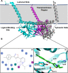
Figure 1
Figure 1
(A) Cartoon representation of the σ1R crystal structure (PDB ID: 5HK1); each color outlines a distinguished σ1R protomer which forms the σ1R trimer. (B) 2D and 3D representation of protein–ligand interactions of the σ1R with PD144418 (PDB ID: 5HK1). The ionic bond between the basic nitrogen of PD144418 and the Glu172 amino acid residue is shown as a dashed yellow line.
Figure 2
Figure 2
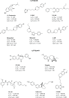
Selected historic and representative σR ligands.
Figure 3
Figure 3
σRs ligands in clinical trials.
Figure 4
Figure 4
(A) The proposed pharmacophore model by Gilligan et al. (B) Glennon’s pharmacophore model.
Figure 5
Figure 5
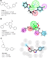
3D pharmacophore models for σ1R: (A) Pharmacophore mapping of compound A in 3D models derived by Vio et al. (B) Pharmacophore mapping of compound B in 3D models derived by Meyer et al. (C) Pharmacophore mapping of PD144418 in 3D models derived by ESTEVE. Color coded as follows: PI (red), HYAr or HYD (light blue), HY (pink), HBA (light green), excluded volumes (gray). Adapted with permission from refs ( and 83). Copyright 2009 and 2012 American Chemical Society.
Figure 6
Figure 6
3D pharmacophore models for σ2R: (A) Pharmacophore mapping of compound C in 3D models derived by Vio et al. (B) Pharmacophore mapping of compound D in 3D models derived by Iyamu et al. Color coded as follows: PI (red), HYAr or HYD (light blue), HY (pink), HBA (light green), excluded volumes (gray).
Figure 7
Figure 7
Schematized representation of the high-throughput structure-based computational docking approach for the discovery of new σ1R ligands proposed by Greenfield et al. Adapted with permission from ref (90). Copyright 2020 American Chemical Society.
Figure 8
Figure 8
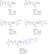
Representative structures of antiproliferative σ1R ligands with their σRs binding profile.
Figure 9
Figure 9
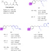
N,N-Dialkyl and N-alkyl-N-aralkyl fenpropimorph-derived compounds 16 and their σRs binding profile.
Figure 10
Figure 10
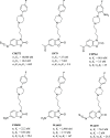
Spipethiane and general structure of spipethiane derivatives 711 and 1215 with their σRs binding profile.
Figure 11
Figure 11
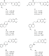
Structures of selected spiropiperidines with a thienofuran and thienopyran scaffold (1621) and their σRs binding profile.
Figure 12
Figure 12
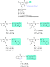
Historically relevant ethylenediamine 2224 with their σRs binding profile. The ethylenediamine structure is highlighted in light blue.
Figure 13
Figure 13
General structures of piperazine derivatives and structures of compounds 2528 with σRs binding profile.
Figure 14
Figure 14
General structure of bicyclic piperazines, chemical structures of 6,8-diazabicyclo[3.2.2]nonanes 29 and 30, and 7,9-diazabicyclo[4.2.2]decane derivatives 3137 with their σRs binding profile.
Figure 15
Figure 15
General structure of 2,5-diazabicyclo[2.2.2]octane derivatives 3843 and ent-3843.
Figure 16
Figure 16
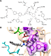
(A) 2D schematic representation of the identified interactions between compound 42 and the main amino acid residues. (B) 3D protein–ligand binding interactions of compound 42 with the σ1R homology model. Color-coded as follows: PI (red), HYAr or HYD (light blue), HY (pink), HBA (light green), π-interactions (Arg119 and Tyr120, cyan), salt bridge (Asp126, red), hydrophobic interactions (Ile128, Phe133, Tyr173, and Leu186, purple), and hydrogen bond (Thr181, green). Adapted with permission from ref (132). Copyright 2016 American Chemical Society.
Figure 17
Figure 17
Representative structures for different chemical classes of σ2R ligands.
Figure 18
Figure 18
Chemical structure and σRs binding profile of selective N-substituted 9-azabicyclo[3.3.1]nonan-3α-yl phenylcarbamate derivatives and conjugated derivative SW III-123.
Figure 19
Figure 19
Early structural modification of siramesine and its analogs 4448.
Figure 20
Figure 20
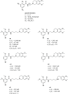
General structure and σRs binding profile of siramesine-related derivatives.
Figure 21
Figure 21
General structure and σRs binding profile of siramesine-related derivatives 51, 52, and 53.
Figure 22
Figure 22
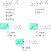
General structure of PB28 analogs with reduced lipophilicity and σRs binding profile of compound 54.
Figure 23
Figure 23
Representative structural modification for PB28 analogs on propylene linker and tetralin scaffold (55 and 56), piperazine ring (PB221), basic N-atom (57), piperazine substitution (58).
Figure 24
Figure 24
Chemical structure and σRs binding profile of N-(4-fluorophenyl)piperazine derivatives.
Figure 25
Figure 25
Chemical structure and σRs binding profile of early 6,7-dimethoxy-1,2,3,4-tetrahydroisoquinoline analogs 5964, reported by Mach and co-workers.
Figure 26
Figure 26
Chemical structure and σRs binding profile of 6,7-dimethoxy-1,2,3,4-tetrahydroisoquinoline analogs 6567.
Figure 27
Figure 27
Chemical structure and σRs binding profile of 6,7-dimethoxy-1,2,3,4-tetrahydroisoquinoline analogs 6870.
Figure 28
Figure 28
Chemical structure and σRs binding profile of compounds 7175.
Figure 29
Figure 29
Chemical structure and σRs binding profile of compounds 7681.
Figure 30
Figure 30
Chemical structures of 3-alkoxyisoxazoles 8290 and their σRs binding profile.

References

    1. Heron M.; Anderson R. N. Changes in the Leading Cause of Death: Recent Patterns in Heart Disease and Cancer Mortality. NCHS data Brief. 2016, 1–8. - PubMed
    1. Chidambaram M.; Manavalan R.; Kathiresan K. Nanotherapeutics to overcome conventional cancer chemotherapy limitations. J. Pharm. Pharm. Sci. 2011, 14, 67–77. 10.18433/J30C7D. - DOI - PubMed
    1. Vanneman M.; Dranoff G. Combining immunotherapy and targeted therapies in cancer treatment. Nat. Rev. Cancer 2012, 12, 237–251. 10.1038/nrc3237. - DOI - PMC - PubMed
    1. Kunick C. Novel molecular targets in cancer chemotherapy waiting for discovery. Curr. Med. Chem.: Anti-Cancer Agents 2004, 4, 421–423. 10.2174/1568011043352858. - DOI - PubMed
    1. Shi Z.; Guo H.-Q.; Cohen P. A.; Yang D.-H. Editorial: Novel Targets and Biomarkers in Solid Tumors. Front. Pharmacol. 2019, 10, 828.10.3389/fphar.2019.00828. - DOI - PMC - PubMed

Publication types

MeSH terms