. 2021 Jun 29;12(3):e0100821.
doi: 10.1128/mBio.01008-21. Epub 2021 May 26.

Dot/Icm-Dependent Restriction of Legionella pneumophila within Neutrophils

Affiliations

Dot/Icm-Dependent Restriction of Legionella pneumophila within Neutrophils

Christopher T D Price et al. mBio. .

Abstract

The Dot/Icm type IV secretion system (T4SS) of Legionella pneumophila is essential for lysosomal evasion and permissiveness of macrophages for intracellular proliferation of the pathogen. In contrast, we show that polymorphonuclear cells (PMNs) respond to a functional Dot/Icm system through rapid restriction of L. pneumophila. Specifically, we show that the L. pneumophila T4SS-injected amylase (LamA) effector catalyzes rapid glycogen degradation in the PMNs cytosol, leading to cytosolic hyperglucose. Neutrophils respond through immunometabolic reprogramming that includes upregulated aerobic glycolysis. The PMNs become activated with spatial generation of intracellular reactive oxygen species within the Legionella-containing phagosome (LCP) and fusion of specific and azurophilic granules to the LCP, leading to rapid restriction of L. pneumophila. We conclude that in contrast to macrophages, PMNs respond to a functional Dot/Icm system, and specifically to the effect of the injected amylase effector, through rapid engagement of major microbicidal processes and rapid restriction of the pathogen. IMPORTANCE Legionella pneumophila is commonly found in aquatic environments and resides within a wide variety of amoebal hosts. Upon aerosol transmission to humans, L. pneumophila invades and replicates with alveolar macrophages, causing pneumonia designated Legionnaires' disease. In addition to alveolar macrophages, neutrophils infiltrate into the lungs of infected patients. Unlike alveolar macrophages, neutrophils restrict and kill L. pneumophila, but the mechanisms were previously unclear. Here, we show that the pathogen secretes an amylase (LamA) enzyme that rapidly breakdowns glycogen stores within neutrophils, and this triggers increased glycolysis. Subsequently, the two major killing mechanisms of neutrophils, granule fusion and production of reactive oxygen species, are activated, resulting in rapid killing of L. pneumophila.

Keywords: granules; phagosome; polymorphonuclear leukocytes; reactive oxygen species.


Figures
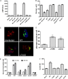
FIG 1
FIG 1
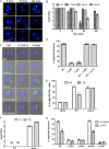
Dot/Icm-injected LamA triggers glycogenolysis in PMNs, resulting in cytosolic hyperglucose and upregulated glycolysis. (A) Adenylate cyclase (Cya) reporter fusion translocation assays of LamA expressed by wild-type L. pneumophila and the translocation-deficient ΔT4SS mutant. The Cya-RalF effector fusion was used a positive control. PMNs were infected for 1 h in triplicate, and cAMP production was assessed by ELISA. To prevent phagocytosis, cytochalasin D was used. Data are shown as mean cAMP concentration ± SD, n = 3. ***, Student's t test of WT-RalF or WT-LamA versus WT-Cya; P < 0.0001. (B) Quantification of cytosolic glycogen concentrations in PMNs starved of glucose or infected wild type, ΔT4SS, ΔlamA or lamA/C and its catalytically inactive mutant, and formalin-killed wild-type bacteria at 1 h postinfection. Data are shown as mean glycogen concentration ± SD; n = 3. **, **, and ***, Student's t test of mock minus glucose cells, WT, or lam/C versus mock-treated cells; P = 0.0037, P = 0.0014, and P = 0.0007, respectively. (C) Representative z-stack confocal microscopy images of PMNs infected with various L. pneumophila strains (green) and glycogen granules were labeled by antibody (red). (D) Quantification of glycogen granules per cell at 15 min postinfection. Glycogen granules were counted in z-stack confocal images, and data points show mean granules/infected cell ± SD (n = 100 infected cells) and are representative of three independent experiments. ***, Student's t test of glycogen granules in either ΔT4SS- or ΔlamA-infected cells versus wild type-infected cells; P < 0.0001. (E) Quantification of cytosolic glucose-6-phosphate levels in PMNs infected with L. pneumophila strains at 15 and 60 min postinfection. Data are shown as mean glucose-6-phosphate concentration ± SD, n = 3. ***, Student's t test of glucose-6-phosphate levels in either wild type- or lamA/C-infected cells versus uninfected cells; P < 0.0001. (F) Lactate levels in cell culture supernatants of PMNs infected for 1 h with wild type, ΔT4SS, ΔlamA, lamA/C, or formalin-killed wild type. Data represent the mean lactate concentration ± SD, n = 3. *** and **, Student's t test of lactate level in the culture supernatant of wild type or lamA/C-infected PMNs versus mock-infected cells; P < 0.001 and P < 0.0013, respectively. Unless otherwise stated, n represents technical replicates, and data shown are representative of three independent biological replicates.
FIG 2
FIG 2
Dot/Icm- and LamA-dependent rapid killing of L. pneumophila by PMNs. (A) Representative confocal images of PMNs infected with either the wild type or the ΔT4SS or ΔlamA mutants at 5, 15, and 60 min postinfection. Bacteria are labeled with an anti-Legionella antibody (green), and nuclei are stained with DAPI (blue). (B) Survival of L. pneumophila in PMNs was determined by measuring recoverable CFU from infected cells. PMNs were infected with the wild type or the ΔT4SS or ΔlamA mutants or the complemented lamA/C mutant, and CFU were determined at 5, 15, 60, and 480 min postinfection. Data are shown as mean log10 CFU ± SD; n = 3. ***, Student's t test CFU at 15 and 60 min relative to 5 min; P < 0.0001. (C and D) Degradation of the ΔT4SS or ΔlamA mutants in PMNs when coinhabiting the same cell with wild-type bacteria. PMNs were infected with the wild type expressing mCherry alone (red), the ΔT4SS or ΔlamA mutants alone, or with wild type coinfected with ΔT4SS or ΔlamA mutants. Following 1 h infection, bacterial morphology was examined by confocal microscopy. Representative confocal images are shown in panel C and quantification in panel D. Data are shown as mean percent degraded bacteria ± SD; n = 100, ***, Student's t test of percent degraded ΔT4SS or ΔlamA mutants in PMNs versus wild-type bacterial cells; P < 0.0001. (E) Analysis of Ly6g+ CD11b+ PMN infiltration in lungs of mock-infected mice or infected with either the wild type or ΔlamA mutant at 4 h and 12 h postinfection. (F) Survival of the wild type or the ΔlamA mutant in Lyg6+ CD11b+ PMNs isolated from the lungs of infected mice. (G) Survival of L. pneumophila in PMNs during inhibition of glycolysis with 2-deoxyglucose was determined by confocal microscopy. PMNs pretreated with 2-deoxyglucose were infected with wild type or the ΔT4SS or ΔlamA mutants or formalin-killed wild type, and bacterial morphology was examined at 15 min postinfection. ***, Student's t test of percent degraded wild type or lamA/C bacteria in PMNs treated with 2-deoxyglucose versus untreated cells; P < 0.0001. Unless otherwise stated, n represents technical replicates, and data shown are representative of three independent biological replicates.
FIG 3
FIG 3
Generation of a robust ROS burst by PMNs in response to cytosolic hyperglucose generated by LamA-mediated glycogenolysis. (A) Determination of ROS produced by PMNs in response to L. pneumophila infection. PMNs were infected with either the wild-type bacteria or the ΔT4SS and ΔlamA mutants, the complemented lamA/C mutant, or formalin-killed wild type for 1 h. Additionally, PMNs were stimulated with zymosan or PMA as a positive control for ROS production. *, Student's t test of ROS production by PMNs infected with either ΔT4SS or the ΔlamA mutant versus wild type infected PMNs; P = 0.0277, 0.0432, respectively. (B) Representative confocal microscopy images showing spatial subcellular localization of ROS production in the pathogen containing phagosome. PMNs treated with NBT were infected with wild type or the ΔT4SS or ΔlamA mutants, the complement lamA/C mutant, or formalin-killed wild type for 1 h. Bacteria were stained using an anti-L. pneumophila antibody (red), and nuclei were stained with DAPI (blue). NBT staining is shown in differential interference contrast (DIC) images as black deposits. (C) The contribution of ROS to rapid bacterial killing was determined using the inhibitor DPI. PMNs were pretreated with DPI and infected with wild-type bacteria, and recoverable CFU were determined at 5 and 15 min postinfection. Data are shown as mean log10 CFU ± SD; n = 3. ***, Student's t test of wild-type CFU in DPI-treated PMNs at 15 min versus untreated PMNs; P < 0.0001. (D) Representative confocal images showing spatial subcellular colocalization of p47phox with the L. pneumophila phagosome. PMNs were infected with wild type or the ΔT4SS or ΔlamA mutants for 15 min. Bacteria are stained with an anti-Legionella antibody (green), and p47phox is stained with anti-p47phox (red). (E) Quantification of p47phox colocalization to the pathogen-containing phagosome at 15 min postinfection. Data are shown as mean percent colocalization ± SD; n = 100. ***, *, and **, Student's t test of percent colocalization of p47phox to phagosomes harboring the ΔT4SS or ΔlamA mutants, formalin-killed wild type, or opsonized S. aureus versus wild type containing phagosomes; P = 0.0008, = 0.0288, and = 0.0043, respectively. Unless otherwise stated, n represents technical replicates, and data shown are representative of three independent biological replicates.
FIG 4
FIG 4
LamA-dependent fusion of specific and azurophilic granules to the L. pneumophila phagosome. (A) Representative confocal images showing colocalization of lactoferrin with the L. pneumophila phagosome. PMNs were infected with wild type or the ΔT4SS or ΔlamA mutants, formalin-killed wild type, or opsonized S. aureus for 15 min. Bacteria are stained with an anti-Legionella antibody (green), and lactoferrin is stained with anti-lactoferrin (red). (B) Quantification of lactoferrin colocalization to the L. pneumophila phagosome at 15 min postinfection. Data are shown as mean percent colocalization ± SD; n = 100. ***, Student's t test of percent colocalization of lactoferrin to phagosomes harboring the ΔT4SS or ΔlamA mutants or formalin-killed wild type versus wild type containing phagosomes; P < 0.0001. (C) Representative confocal images showing colocalization of elastase with the L. pneumophila phagosome. PMNs were infected with wild type or the ΔT4SS or ΔlamA mutants, formalin-killed wild type, or opsonized S. aureus for 15 min. Bacteria were labeled with an anti-Legionella antibody (green), and elastase is labeled with anti-elastase antibodies (red). (D) Quantification of elastase colocalization to the L. pneumophila phagosome at 15 min postinfection. Data are shown as mean percent colocalization ± SD; n = 100. ***, Student's t test of percent colocalization of elastase to phagosomes harboring the ΔT4SS or ΔlamA mutants or formalin-killed wild type versus wild type containing phagosomes; P < 0.0001. (E) Relative contribution of specific and azurophilic granules to rapid bacterial killing was determined using a pan-protease, cathepsin G, and elastase inhibitors. PMNs were pretreated with DPI and infected with wild-type bacteria, and recoverable CFU were determined at 5 and 15 min postinfection. Data are shown as mean log10 CFU ± SD; n = 3. ***, Student's t test of wild-type CFU in PMNs treated with the pan-protease or elastase inhibitors at 15 min versus untreated PMNs; P < 0.0001. Unless otherwise stated, n represents technical replicates, and data shown are representative of three independent biological replicates.

Similar articles

Cited by

References

    1. Fields BS. 1996. The molecular ecology of legionellae. Trends Microbiol 4:286–290. doi:10.1016/0966-842x(96)10041-x. - DOI - PubMed
    1. Molmeret M, Horn M, Wagner M, Santic M, Abu Kwaik Y. 2005. Amoebae as training grounds for intracellular bacterial pathogens. Appl Environ Microbiol 71:20–28. doi:10.1128/AEM.71.1.20-28.2005. - DOI - PMC - PubMed
    1. Harb OS, Gao L-Y, Abu Kwaik Y. 2000. From protozoa to mammalian cells: a new paradigm in the life cycle of intracellular bacterial pathogens. Environ Microbiol 2:251–265. doi:10.1046/j.1462-2920.2000.00112.x. - DOI - PubMed
    1. Desai SK, Kenney LJ. 2019. Switching lifestyles is an in vivo adaptive strategy of bacterial pathogens. Front Cell Infect Microbiol 9:421. doi:10.3389/fcimb.2019.00421. - DOI - PMC - PubMed
    1. Shuman HA, Purcell M, Segal G, Hales L, Wiater LA. 1998. Intracellular multiplication of Legionella pneumophila: human pathogen or accidental tourist? Curr Top Microbiol Immunol 225:99–112. doi:10.1007/978-3-642-80451-9_6. - DOI - PubMed

Publication types

MeSH terms

LinkOut - more resources