. 2021 Aug 10;95(17):e0051821.
doi: 10.1128/JVI.00518-21. Epub 2021 Aug 10.

Identification of an Intramolecular Switch That Controls the Interaction of Helicase nsp10 with Membrane-Associated nsp12 of Porcine Reproductive and Respiratory Syndrome Virus

Affiliations

Identification of an Intramolecular Switch That Controls the Interaction of Helicase nsp10 with Membrane-Associated nsp12 of Porcine Reproductive and Respiratory Syndrome Virus

Yunhao Hu et al. J Virol. .

Abstract

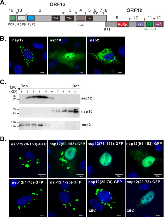
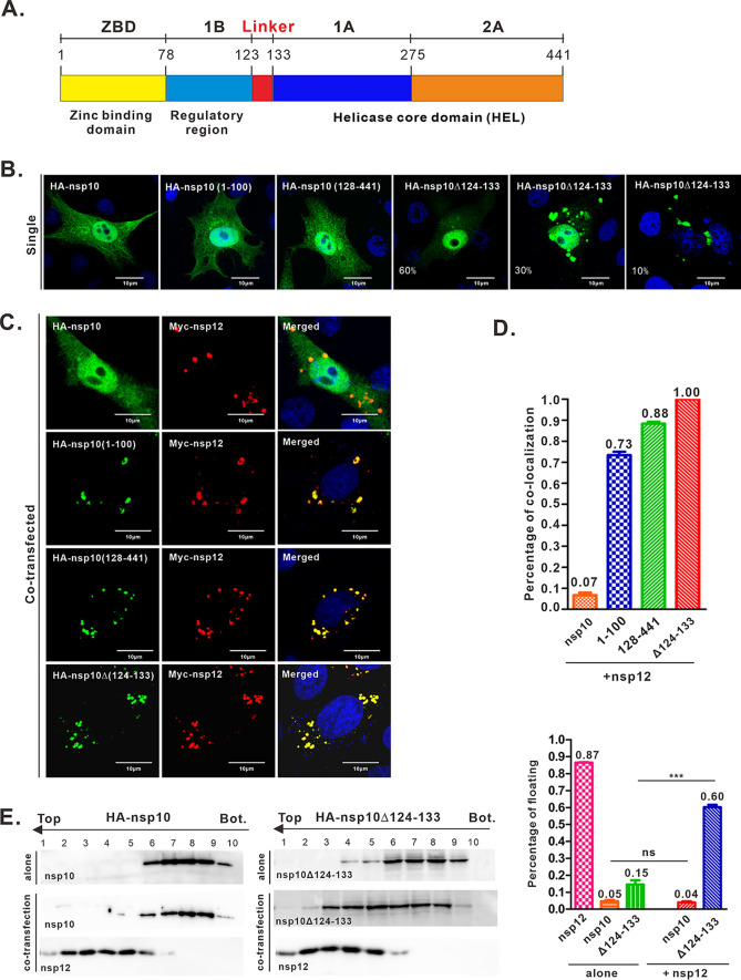
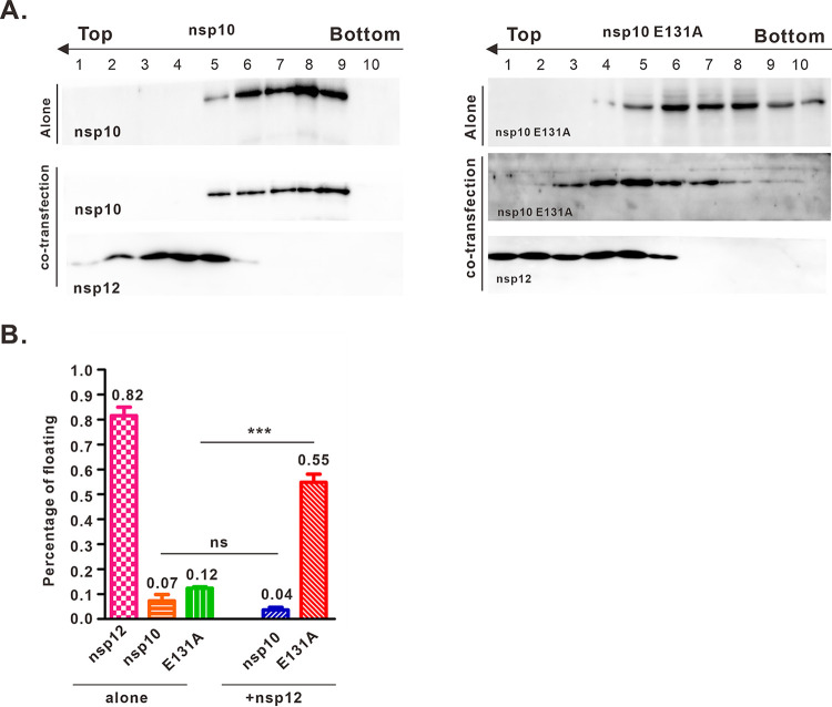
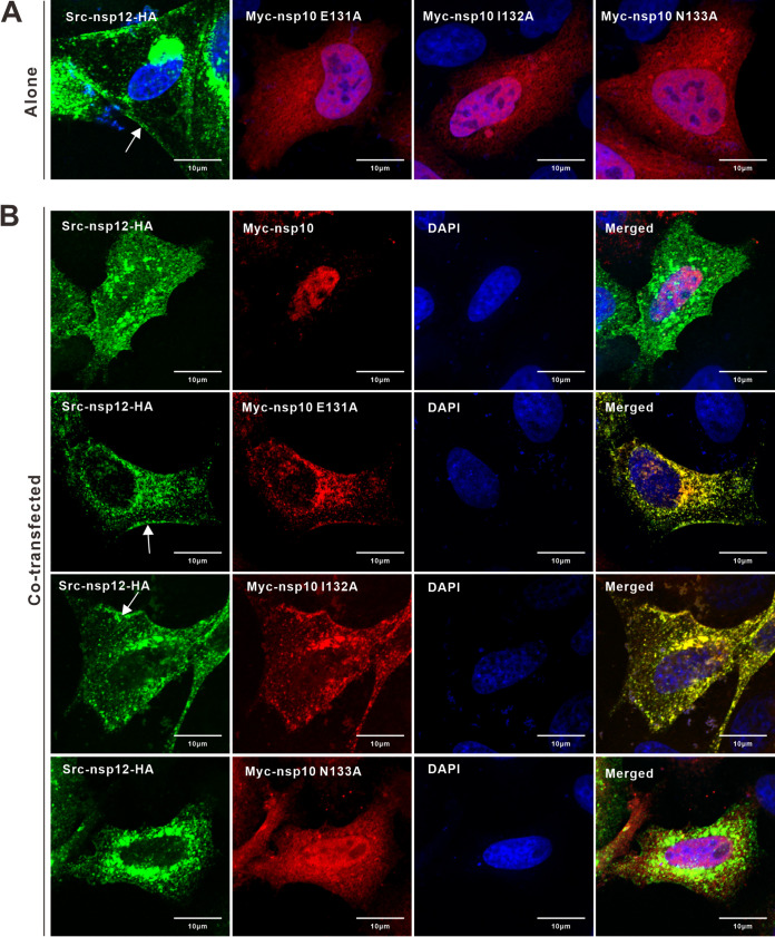
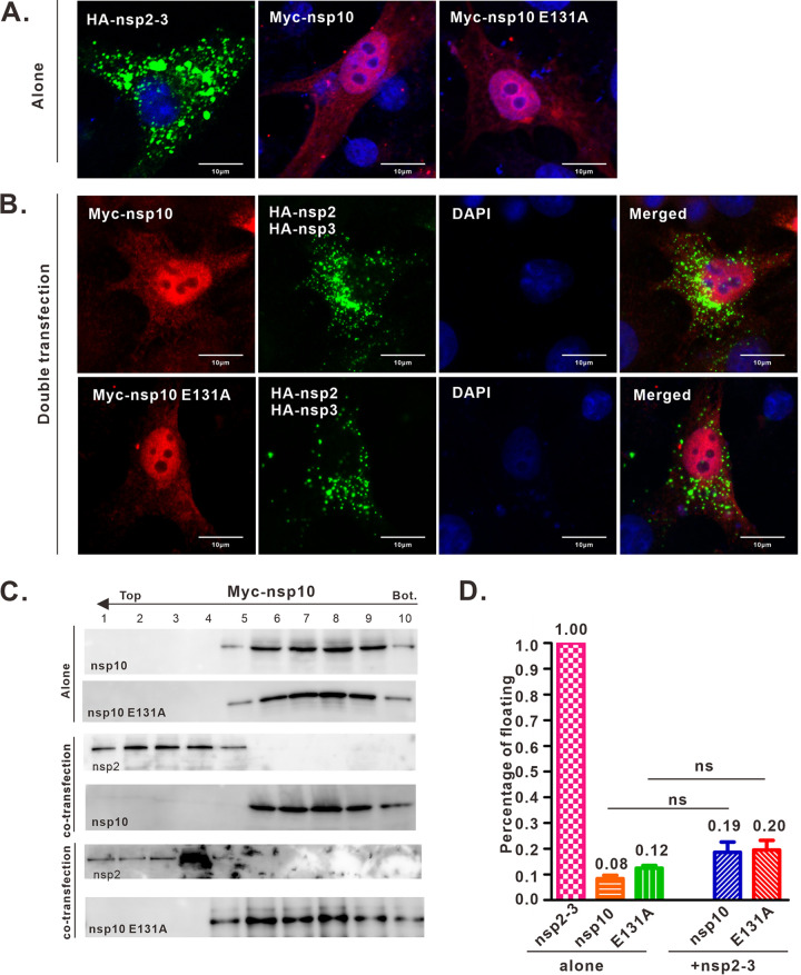
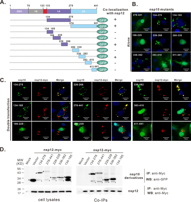
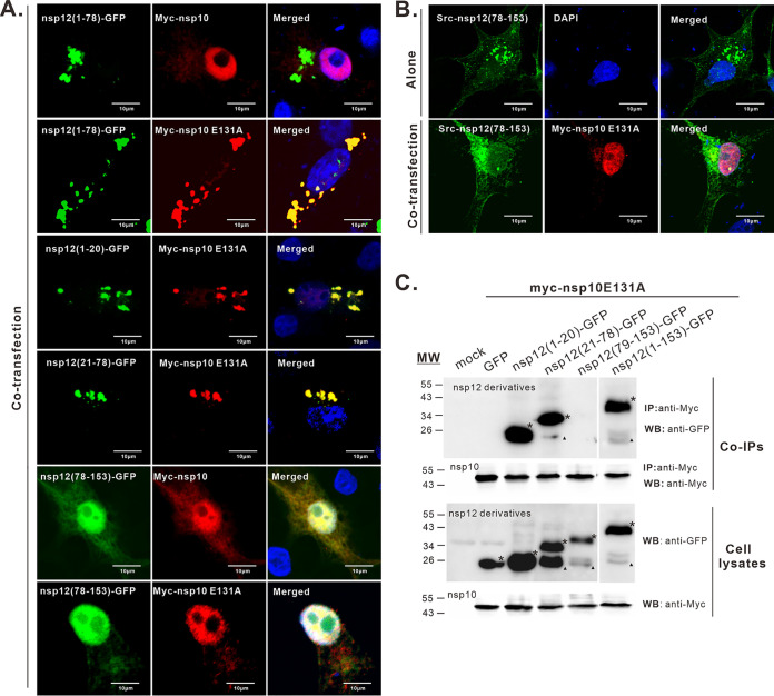
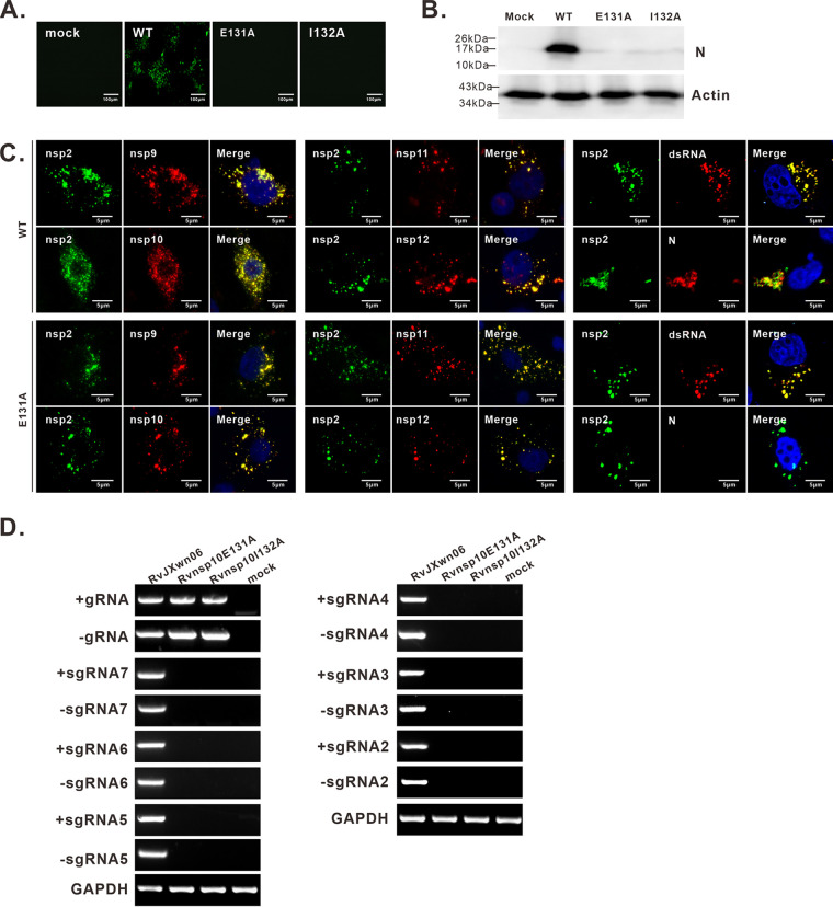
A critical step in replication of positive-stranded RNA viruses is the assembly of replication and transcription complexes (RTC). We have recently mapped the nonstructural protein (nsp) interaction network of porcine reproductive and respiratory syndrome virus (PRRSV) and provided evidence by truncation mutagenesis that the recruitment of viral core replicase enzymes (nsp9 and nsp10) to membrane proteins (nsp2, nsp3, nsp5, and nsp12) is subject to regulation. Here, we went further to discover an intramolecular switch within the helicase nsp10 that controls its interaction with the membrane-associated protein nsp12. Deletion of nsp10 linker region amino acids 124 to 133, connecting domain 1B to 1A, led to complete relocalization and colocalization in the cells coexpressing nsp12. Moreover, single-amino-acid substitutions (e.g., nsp10 E131A and I132A) were sufficient to enable the nsp10-nsp12 interaction. Further proof came from membrane floatation assays that revealed a clear movement of nsp10 mutants, but not wild-type nsp10, toward the top of sucrose gradients in the presence of nsp12. Interestingly, the same mutations were not able to activate the nsp10-nsp2/3 interaction, suggesting a differential requirement for conformation. Reverse genetics analysis showed that PRRSV mutants carrying the single substitutions were not viable and were defective in subgenomic RNA (sgRNA) accumulation. Together, our results provide strong evidence for a regulated interaction between nsp10 and nsp12 and suggest an essential role for an orchestrated RTC assembly in sgRNA synthesis. IMPORTANCE Assembly of replication and transcription complexes (RTC) is a limiting step for viral RNA synthesis. The PRRSV RTC macromolecular complexes are comprised of mainly viral nonstructural replicase proteins (nsps), but how they come together remains elusive. We previously showed that viral helicase nsp10 interacts nsp12 in a regulated manner by truncation mutagenesis. Here, we revealed that the interaction is controlled by single residues within the domain linker region of nsp10. Moreover, the activation mutations lead to defects in viral sgRNA synthesis. Our results provide important insight into the mechanisms of PRRSV RTC assembly and regulation of viral sgRNA synthesis.

Keywords: assembly; nonstructural protein; porcine reproductive and respiratory syndrome virus; regulated interaction; replication and transcription complexes; subgenomic RNA synthesis.


Figures

FIG 1
FIG 1
PRRSV nsp12 is a membrane-associated protein. (A) Diagram of PRRSV ORF1a and ORF1b, which are posttranslationally cleaved by virus-encoded papain-like proteases (PCP1а and PCP1β), cysteine protease (PLP2), and 3C-like serine protease (3CL). nsp2, nsp3, and nsp5 are proteins that contain transmembrane (TM) domains. The following signature regions are indicated: ribosomal frameshift signal (RFS), RNA-dependent RNA-polymerase (RdRp), helicase (HEL), endoribonucleases (NendoU), and arterivirus-specific domain (AsD). (B) Cellular localization of Myc-tagged nsp12 and nsp10 in transfected BHK21 cells probed with antibodies to the Myc epitope. (C) Membrane flotation analyses of nsp12 and nsp10. HEK-293T cells were transfected with Myc-nsp10 or Myc-nsp12. At 16 to 20 h posttransfection, the cells were osmotically disrupted, and the ability of each protein to float to the upper fractions of sucrose step gradients was examined. Ten equal fractions were collected, and proteins were analyzed by Western blotting with rabbit anti-Myc pAb. The tops and bottoms (Bot.) of the gradients are indicated. MW, molecular weight. (D) Subcellular localization of nsp12 and its truncations. Expression plasmids carrying the genes for nsp12 derivatives were transfected into BHK-21 cells. At 18 to 24 h posttransfection, the cells were fixed and nsp12 constructs were detected by the fluorescence of their GFP tags (green). The cell nuclei were stained with DAPI (blue). The representative images were captured with a Nikon confocal microscope and processed using Image J. Oil objective, 100×; zoom, 1×.
FIG 2
FIG 2
Deletion of the nsp10 linker region activates its interaction with nsp12. (A) The domain organization of PRRSV nsp10. ZBD, Zn-binding domain; HEL, helicase core domain; 1B, regulatory domain; 1A, RecA-like domain 1A; 2A, RecA-like domain 2A. (B) HeLa cells were singly transfected to express HA-nsp10 and its truncation mutants. At 18 to 24 h posttransfection, the cells were fixed and stained with monoclonal antibodies to HA epitope. (C) Colocalization analysis of nsp12 with nsp10 and its derivatives. HeLa cells were transfected to express the indicated protein pairs and stained with antibodies to both HA and myc epitopes at 18 to 24 h posttransfection. The representative images were captured with a Nikon confocal microscope and processed using Image J. Oil objective, 100×; zoom, 1×. (D) Quantitative analysis for colocalization relationships in cells coexpressing nsp12 with nsp10 or its derivatives. The percentages of cells showing colocalization were determined. (E) Membrane flotation analyses of nsp10 or its derivative in the presence or absence of nsp12. Quantitative analysis was determined by immunoblots from three independent experiments. The results are shown as the percentage of floating protein (top five fractions) relative to the total protein (all fractions) (averages are shown on top of each bar). ***, P < 0.001; ns, not significant. The error bars indicate standard deviations.
FIG 3
FIG 3
Identification of key residues activating nsp10-nsp12 interaction. (A) Sequence information for nsp10 linker domain and the strategy for constructing nsp10 mutants. The nsp12-nsp10 binding activity is summarized on the right. +, interaction; −, no interaction. (B) Subcellular localization of HA-nsp10 mutants in singly transfected HeLa cells. (C) Colocalization analysis of nsp10 mutants and nsp12 in transfected HeLa cells. At 18 to 24 h posttransfection, the cells were stained with the antibodies specific for Myc tag (red) and HA tag (green). The representative images were captured with a Nikon confocal microscope and processed using Image J. Oil objective, 100×; zoom, 1×.
FIG 4
FIG 4
Membrane flotation analyses of nsp10 derivatives in the presence or absence of nsp12. (A) HEK-293T cells were transfected with the indicated nsp10 or nsp10 E131A, either alone or with myc-nsp12. At 16 to 20 h posttransfection, the cells were osmotically disrupted, and the ability of each protein to float to the upper fractions of sucrose step gradients was examined. Ten equal fractions were collected, and proteins were analyzed by Western blotting with the mouse anti-HA (1:5,000) and rabbit anti-Myc (1:1,000). The tops and bottoms (Bot.) of the gradients are indicated. (B) Quantitative analysis was determined by immunoblots from three independent experiments. The results are shown as the percentage of floating protein (top five fractions) relative to the total protein (all fractions). ***, P < 0.001; ns, not significant. The error bars indicate standard deviations.
FIG 5
FIG 5
Relocalization analysis of the nsp10-nsp12 interaction. (A) Subcellular localization analysis of Src-nsp12-HA or Myc-tagged nsp10 mutants in singly transfected HeLa cells. (B) Relocalization analysis of nsp10 and its mutants. The indicated derivatives of nsp10 were coexpressed with Src-nsp12 in HeLa cells. Src-nsp12 was detected by anti-HA pAb (green), whereas nsp10 was detected with anti-Myc MAb (red). DAPI was used to stain nuclei (blue). The representative images were captured with a Nikon confocal microscope and processed using Image J. Oil objective, 100×; zoom, 1×.
FIG 6
FIG 6
Conformation required for interaction with nsp12 is different from that for nsp2/3. (A) Cells were singly transfected with HA-nsp2-3, Myc-nsp10, or Myc-nsp10 E131A to show the sites where these proteins accumulate on their own. (B) Myc-nsp10 or Myc-nsp10 E131A was coexpressed with HA-nsp2-3 to look for changes in subcellular localization. Myc-nsp10 and Myc-nsp10 E131A were revealed by a monoclonal antibody specific for the Myc peptide (red). The position of nsp2-3 was revealed by a monoclonal antibody specific for the HA peptide (green). The representative images were captured with a Nikon confocal microscope and processed using Image J. Oil objective, 100×; zoom, 1×. (C) Membrane flotation analyses of nsp10 and nsp10 E131A in the presence or absence of nsp2-3 in transfected HEK-293T cells. At 20 h posttransfection, the cells were osmotically disrupted, and the ability of each protein to float to the upper fractions of sucrose step gradients was examined. Ten equal fractions were collected, and proteins were analyzed by Western blotting with nsp2 pAb to detect nsp2-3 and Myc MAb to detect nsp10 constructs. The tops and bottoms of the gradients are indicated. Quantitative analysis was determined by immunoblots from three independent experiments. The results are shown as the percentage of floating protein (top five fractions) relative to the total protein (all fractions). ***, P < 0.001; ns, not significant. The error bars indicate standard deviations.
FIG 7
FIG 7
Mapping the interaction sites within nsp10 for nsp12. (A) Schematic presentation of PRRSV nsp10 truncations fused to GFP. Their colocalization relationship with nsp12 is summarized on the right, labeled + or −. (B) Subcellular localization of the nsp10 mutants in transfected BHK-21 cells. (C) Colocalization analysis of nsp12 and nsp10 mutants in the cotransfected BHK-21 cells. The representative images were captured with a Nikon confocal microscope and processed using Image J. Oil objective, 100×; zoom, 1×. (D) Co-IP analysis of the interaction between the nsp10 mutants and nsp12. HEK-293T cells were cotransfected to express the indicated protein pairs. At 24 h after transfection, cell lysates were prepared and analyzed by immunoblotting to measure the input expression levels or subjected to co-IP analysis with antibodies to myc. The bound proteins were subjected to Western blotting with a rabbit anti-GFP pAb to reveal the presence of nsp10 truncations. The asterisk indicates the specific signals, while the triangle indicates the likely nonspecific signals.
FIG 8
FIG 8
Mapping the interaction site within nsp12 for nsp10. (A) Colocalization analysis of nsp12 mutants with nsp10 or nsp10 E131A in cotransfected BHK-21 cells. At 18 to 24 h posttransfection, nsp10 and nsp10 E131A were revealed by a monoclonal antibody specific for the Myc epitope (red), whereas nsp12 constructs were detected by the fluorescence of GFP tag (green). (B) Relocalization analysis of the interaction between nsp10 E131A and Src-nsp12 (79-153). Src-tagged nsp12 (79-153) was expressed alone or coexpressed to look for the localization change of nsp10 E131A. The representative images were captured with a Nikon confocal microscope and processed using Image J. Oil objective, 100×; zoom, 1×. (C) Co-IP analysis of the interaction between nsp10 E131A and the GFP-tagged nsp12 mutants. The antibodies used for IP and Western blotting are indicated. The asterisk indicates the specific band, while the triangle indicates the likely nonspecific signals.
FIG 9
FIG 9
Activating mutation is lethal to PRRSV. (A and B) The viability of mutant viruses was assessed in MARC-145 cells by IFA (A) and Western blotting (B) with mouse monoclonal antibodies to N protein. (C) Colocalization analysis of viral replicase proteins, N, and double-stranded RNAs with nsp2 in BHK-21 cells transfected with the infectious cDNA clone plasmid for nsp10 E131A or WT JXwn06. The cells were stained with specific proteins for viral nsp, N, and dsRNA at 48 h posttransfection. The representative images were captured with a Nikon confocal microscope and processed using Image J. Oil objective, 100×; zoom, 1×. (D) A strand-specific PCR method was used to detect positive-strand RNA (+gRNA), negative-strand genomic RNA (−gRNA), positive-strand subgenomic RNA (+sg RNA), and negative-strand subgenomic RNA (−sg RNA) at 48 h posttransfection from BHK-21 cells mock transfected or transfected with the cDNA clone plasmids for WT, nsp10 E131A, and nsp10 I132A.

Similar articles

Cited by

References

    1. Snijder EJ, Decroly E, Ziebuhr J. 2016. The nonstructural proteins directing coronavirus RNA synthesis and processing. Adv Virus Res 96:59–126. 10.1016/bs.aivir.2016.08.008. - DOI - PMC - PubMed
    1. Yan L, Zhang Y, Ge J, Zheng L, Gao Y, Wang T, Jia Z, Wang H, Huang Y, Li M, Wang Q, Rao Z, Lou Z. 2020. Architecture of a SARS-CoV-2 mini replication and transcription complex. Nat Commun 11:5874. 10.1038/s41467-020-19770-1. - DOI - PMC - PubMed
    1. Athmer J, Fehr AR, Grunewald M, Smith EC, Denison MR, Perlman S. 2017. In situ tagged nsp15 reveals interactions with coronavirus replication/transcription complex-associated proteins. mBio 8:e02320-16. 10.1128/mBio.02320-16. - DOI - PMC - PubMed
    1. van Hemert MJ, van den Worm SH, Knoops K, Mommaas AM, Gorbalenya AE, Snijder EJ. 2008. SARS-coronavirus replication/transcription complexes are membrane-protected and need a host factor for activity in vitro. PLoS Pathog 4:e1000054. 10.1371/journal.ppat.1000054. - DOI - PMC - PubMed
    1. Ziebuhr J, Snijder EJ, Gorbalenya AE. 2000. Virus-encoded proteinases and proteolytic processing in the Nidovirales. J Gen Virol 81:853–879. 10.1099/0022-1317-81-4-853. - DOI - PubMed

Publication types

MeSH terms

Substances

LinkOut - more resources