. 2021 Jul 26;95(16):e0083621.
doi: 10.1128/JVI.00836-21. Epub 2021 Jul 26.

NAP1L1 and NAP1L4 Binding to Hypervariable Domain of Chikungunya Virus nsP3 Protein Is Bivalent and Requires Phosphorylation

Affiliations

NAP1L1 and NAP1L4 Binding to Hypervariable Domain of Chikungunya Virus nsP3 Protein Is Bivalent and Requires Phosphorylation

Francisco Dominguez et al. J Virol. .

Abstract

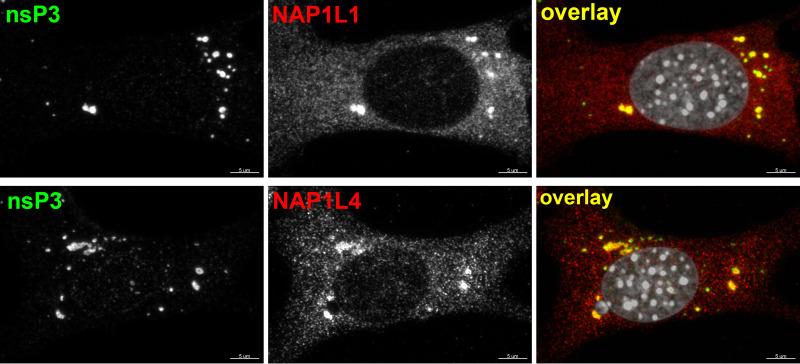
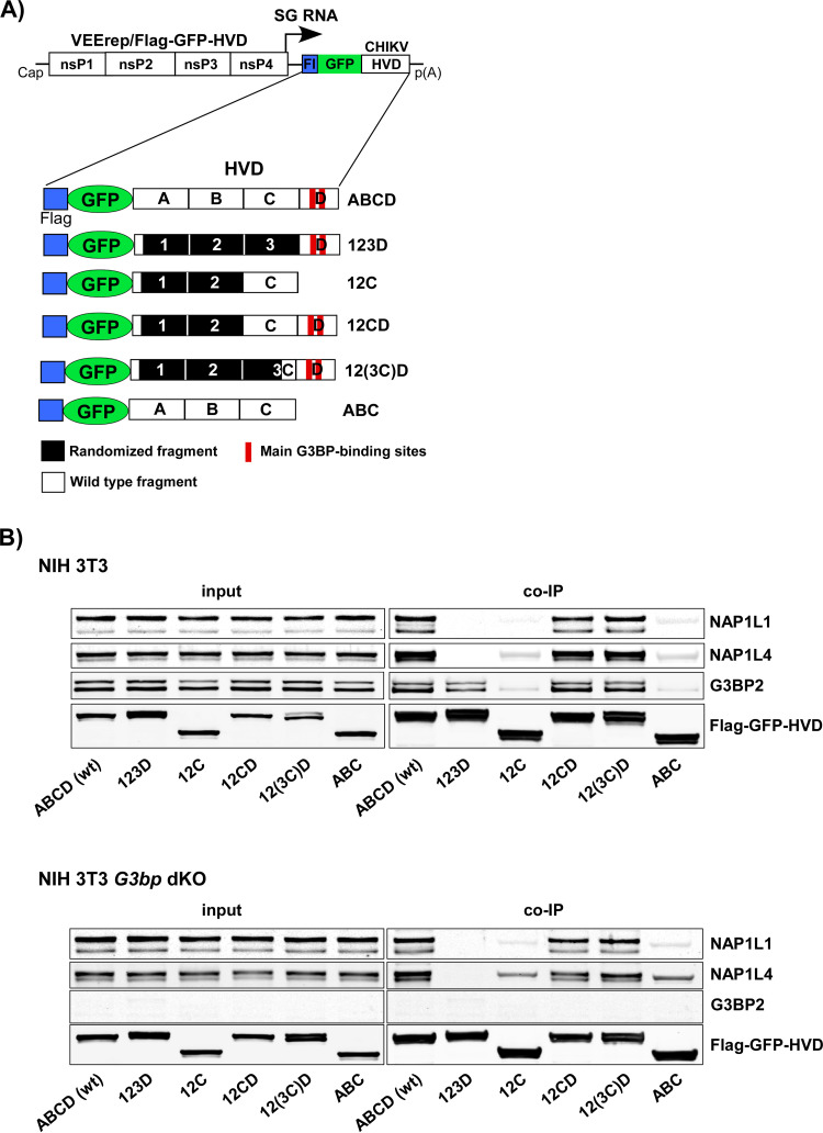
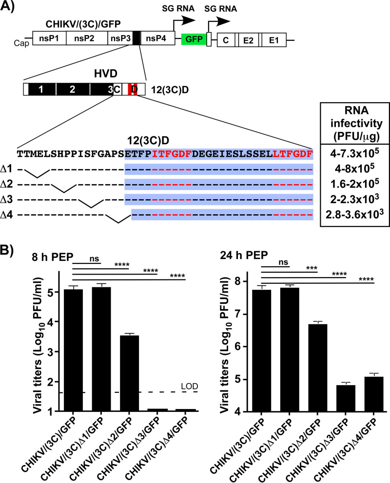
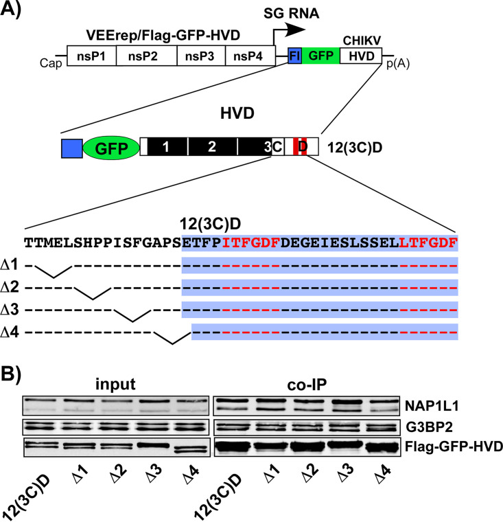
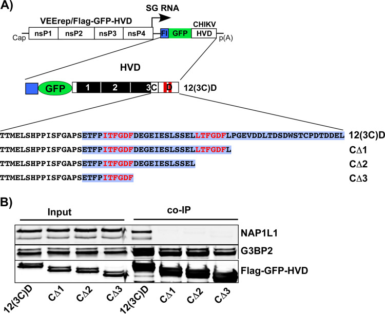
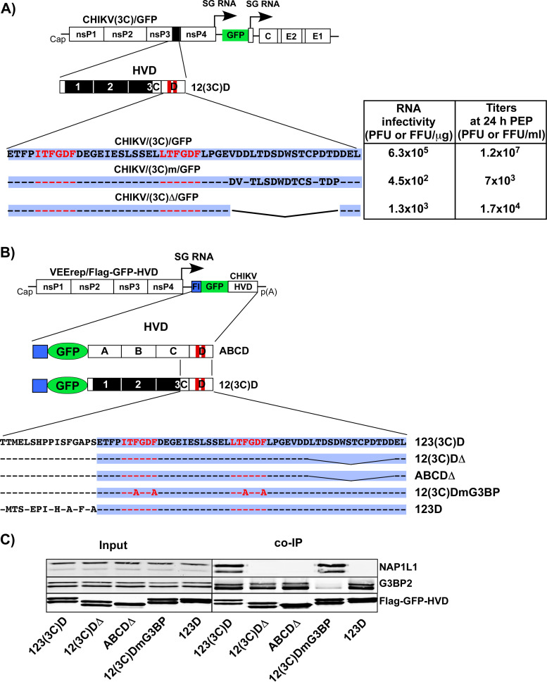
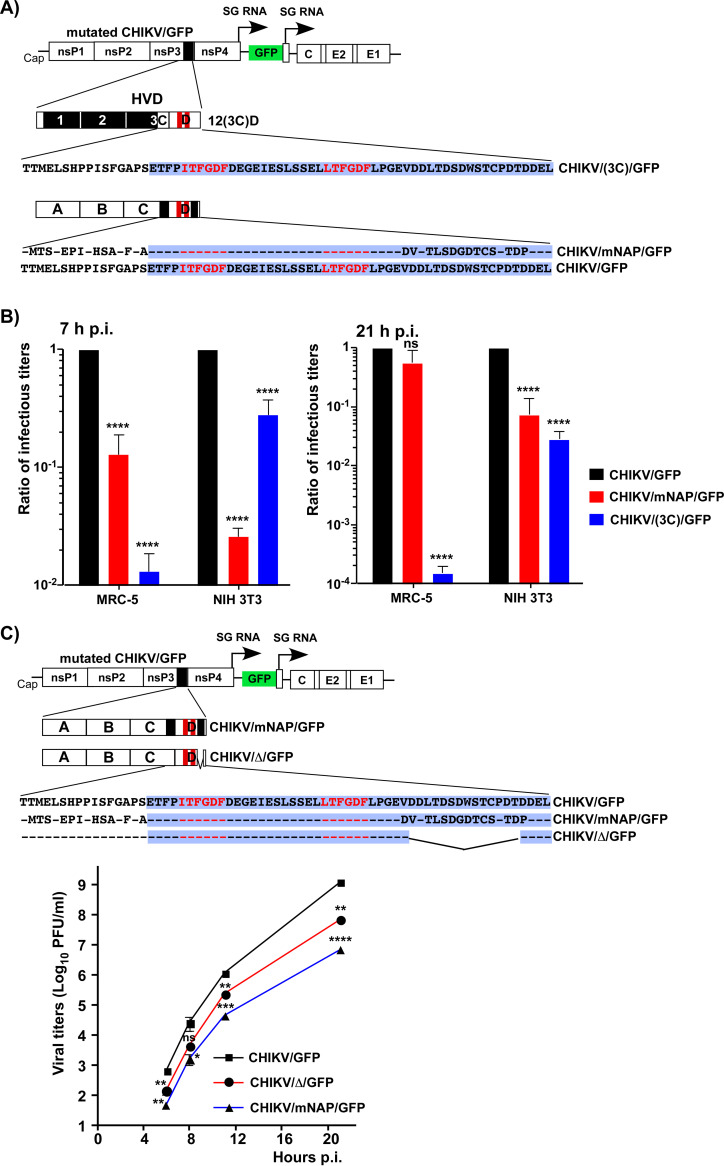
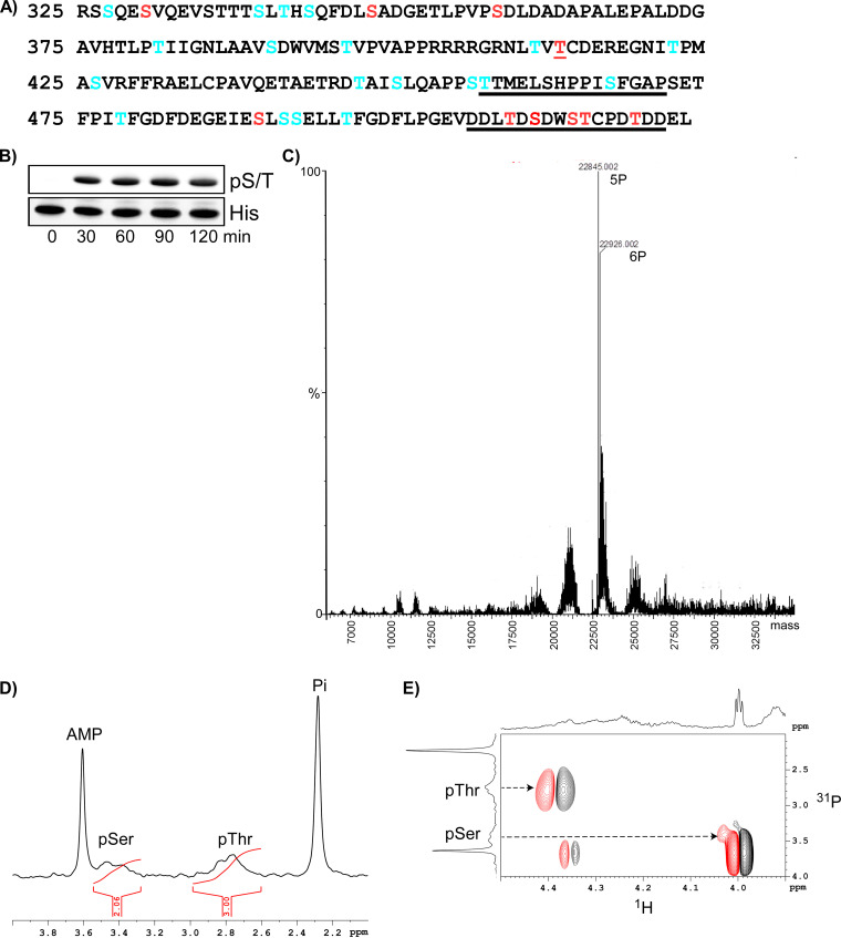
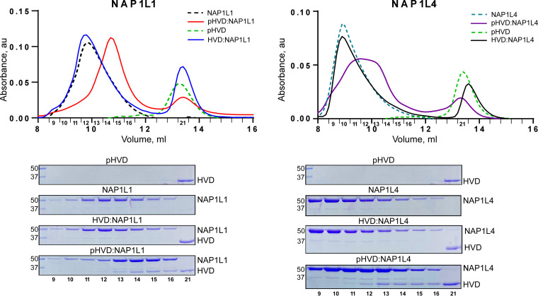
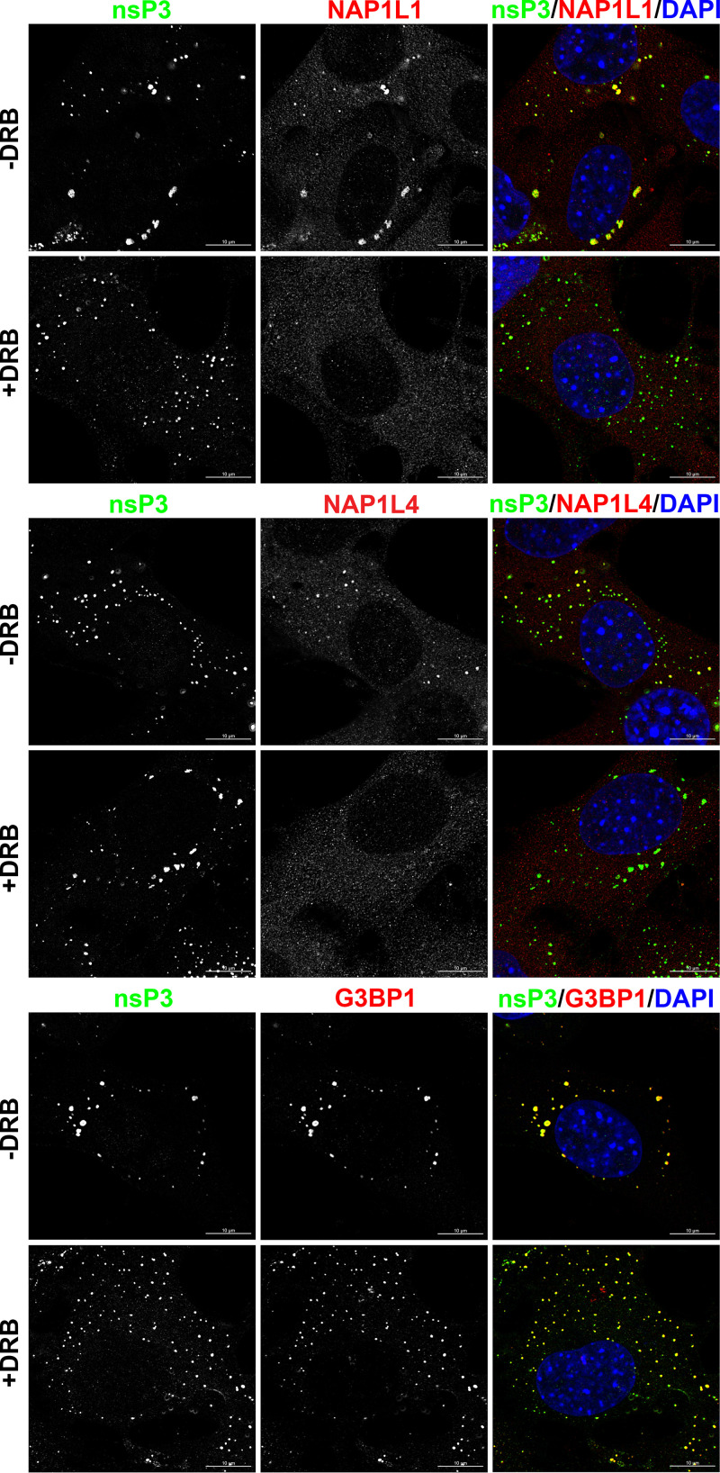
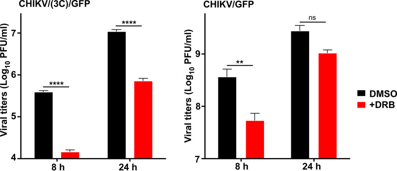
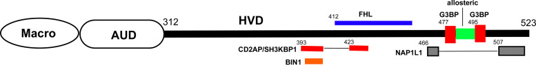
Chikungunya virus (CHIKV) is one of the most pathogenic members of the Alphavirus genus in the Togaviridae family. Within the last 2 decades, CHIKV has expanded its presence to both hemispheres and is currently circulating in both Old and New Worlds. Despite the severity and persistence of the arthritis it causes in humans, no approved vaccines or therapeutic means have been developed for CHIKV infection. Replication of alphaviruses, including CHIKV, is determined not only by their nonstructural proteins but also by a wide range of host factors, which are indispensable components of viral replication complexes (vRCs). Alphavirus nsP3s contain hypervariable domains (HVDs), which encode multiple motifs that drive recruitment of cell- and virus-specific host proteins into vRCs. Our previous data suggested that NAP1 family members are a group of host factors that may interact with CHIKV nsP3 HVD. In this study, we performed a detailed investigation of the NAP1 function in CHIKV replication in vertebrate cells. Our data demonstrate that (i) the NAP1-HVD interactions have strong stimulatory effects on CHIKV replication, (ii) both NAP1L1 and NAP1L4 interact with the CHIKV HVD, (iii) NAP1 family members interact with two motifs, which are located upstream and downstream of the G3BP-binding motifs of CHIKV HVD, (iv) NAP1 proteins interact only with a phosphorylated form of CHIKV HVD, and HVD phosphorylation is mediated by CK2 kinase, and (v) NAP1 and other families of host factors redundantly promote CHIKV replication and their bindings have additive stimulatory effects on viral replication. IMPORTANCE Cellular proteins play critical roles in the assembly of alphavirus replication complexes (vRCs). Their recruitment is determined by the viral nonstructural protein 3 (nsP3). This protein contains a long, disordered hypervariable domain (HVD), which encodes virus-specific combinations of short linear motifs interacting with host factors during vRC assembly. Our study defined the binding mechanism of NAP1 family members to CHIKV HVD and demonstrated a stimulatory effect of this interaction on viral replication. We show that interaction with NAP1L1 is mediated by two HVD motifs and requires phosphorylation of HVD by CK2 kinase. Based on the accumulated data, we present a map of the binding motifs of the critical host factors currently known to interact with CHIKV HVD. It can be used to manipulate cell specificity of viral replication and pathogenesis, and to develop a new generation of vaccine candidates.

Keywords: CK2 kinase; NAP1L1; NAP1L4; alphavirus; chikungunya virus; intrinsically disordered proteins; nsP3; protein phosphorylation; viral pathogenesis; viral replication.


Figures

FIG 1
FIG 1
NAP1L1 and NAP1L4 accumulate in CHIKV nsP3 complexes. NIH 3T3 cells were infected with CHIKV 181/25 at an MOI of 10 PFU/cell in 8-well Ibidi plates. At 7 h p.i., the cells were fixed and stained with CHIKV nsP3- and NAP1-specific Abs and DAPI as described in Materials and Methods. Images were acquired on a Zeiss LSM 800 confocal microscope in Airyscan mode with a 63 × 1.4 NA PlanApochromat oil objective.
FIG 2
FIG 2
NAP1 family members are capable of interacting with the C-terminal fragment of CHIKV HVD independently of G3BP. (A) The schematic presentation of VEEV replicons encoding different variants of CHIKV HVD fused with Flag-GFP. Labels A, B, C, and D indicate wt aa sequences. Fragments 1, 2, and 3 have the aa sequences of the corresponding A, B, and C fragments randomized. In fragment (3C), only the C-terminal 16-aa peptide had the wt sequence. wt and mutated fragments are additionally indicated by open and black boxes, respectively. (B) NIH 3T3 cells and their G3bp dKO derivatives were infected at an MOI of 10 inf.u/cell with the packaged VEEV replicons expressing the indicated fragments of CHIKV HVD. Cells were collected at 4 h p.i. Protein complexes were isolated on magnetic beads as described in Materials and Methods and analyzed by Western blotting with NAP1L1-, NAP1L4-, G3BP2-, and Flag-specific Abs and corresponding secondary Abs. Membranes were scanned on a Li-Cor imager. The experiment was repeated three times with similar results.
FIG 3
FIG 3
Mutations in NAP1-binding site affect CHIKV replication. (A) Schematic presentation of the CHIKV/(3C)/GFP genome, deletions introduced into (3C) fragment, and infectivities of the in vitro-synthesized RNA in the ICA (see Materials and Methods for details). Dashes indicate aa identical to those in HVD of the parental virus. Fragment D is additionally indicated by blue background. (B) BHK-21 cells were electroporated with 3 µg of the in vitro-synthesized RNAs of parental CHIKV/(3C)/GFP and its deletion mutants. Samples of the media were collected at the indicated times postelectroporation, and titers were determined by plaque assay on BHK-21 cells. The ICA and titers of the released viruses were assessed in the same experiment, which was reproducibly repeated three times. Means and standard deviations (SD) are indicated. The significance of differences was determined by one-way analysis of variance (ANOVA) with the Tukey test (***, P < 0.001; ****, P < 0.0001; n= 3).
FIG 4
FIG 4
Adaptive mutations in the macro domain and AUD compensate negative effects of the deletions in the potential NAP1-binding site. (A) The schematic presentation of CHIKV/(3C)/GFP genome and deletions made in potential NAP1-binding site. (B) Schematic presentation of the domain structure of CHIKV nsP3 in the 12(3C)D variant, positions of the deletions introduced upstream of the G3BP-binding site, and adaptive mutations in the macro domain and AUD (indicated by red arrows). Infectivities of the in vitro-synthesized RNAs of the parental deletion mutants and those with adaptive mutations in the ICA (see Materials and Methods for details). The experiment was reproducibly repeated twice, and the results of one experiment are presented. (C) Replication of the variants with adaptive mutations in structured domains and the parental deletion mutants in BHK-21 cells after electroporation of the in vitro-synthesized RNAs. Samples of the media were collected at the indicated times postelectroporation, and titers were determined by plaque assay on BHK-21 cells. The experiment was reproducibly repeated three times. Means and SD are indicated. The significance of differences was determined by one-way ANOVA with the Tukey test (****, P < 0.0001; n= 3).
FIG 5
FIG 5
Deletions in putative NAP1-binding site do not abrogate 12(3C)D HVD interaction with NAP1L1. (A) Schematic presentation of VEEV replicon encoding Flag-GFP-12(3C)D HVD fusion with the indicated deletions in the motif located upstream of the G3BP-binding site. (B) NIH 3T3 cells were infected with packaged replicons at an MOI of 20 inf.u/cell. Cells were harvested at ∼3 h p.i. Protein complexes were isolated on magnetic beads as described in Materials and Methods. They were analyzed by Western blotting with NAP1L1- and Flag-specific Abs and corresponding secondary Abs. Membranes were scanned on a Li-Cor imager. The experiment was repeated twice with identical results. The results of one experiment are presented.
FIG 6
FIG 6
Deletions in the C-terminal fragment of 12(3C)D HVD abrogate its binding of NAP1L1, but not of G3BP. (A) Schematic presentation of VEEV replicons encoding deletion variants of CHIKV 12(3C)D HVD fused with Flag-GFP. Fragment D is additionally indicated by blue background. (B) NIH 3T3 cells were infected with packaged replicons at an MOI of 20 inf.u/cell. Cells were harvested at 3 h p.i. Protein complexes were isolated on magnetic beads and analyzed by Western blotting with NAP1L1-, G3BP2-, and Flag-specific Abs and corresponding secondary Abs. Membranes were scanned on a Li-Cor imager. The experiment was repeated twice with identical results. The results of one experiment are presented.
FIG 7
FIG 7
Modifications in the C-terminal fragment of CHIKV/(3C)/GFP have deleterious effects on viral replication. (A) Schematic presentation of the genomes and HVDs of the designed viruses and aa sequences of their D fragments. Dashes indicate aa identical to those in the wt sequence. The table presents the infectivities of the in vitro-synthesized RNAs in the ICA and viral titers at 24 h postelectroporation. RNA infectivities and titers were measured in PFU and GFP-positive focus-forming units (FFUs) for CHIKV/(3C)/GFP and its derivatives, respectively. The experiment was repeated twice with reproducible results. The data from one experiment are presented. (B) Schematic presentation of VEEV replicons encoding different variants of CHIKV HVD fused with Flag-GFP. Fragment D is additionally indicated by blue background. (C) NIH 3T3 cells were infected with packaged replicons at an MOI of 20 inf.u/cell. Cells were harvested at 3 h p.i. Protein complexes were isolated on magnetic beads and analyzed by Western blotting with NAP1L1-, G3BP2-, and Flag-specific Abs and corresponding secondary Abs. Membranes were scanned on a Li-Cor imager.
FIG 8
FIG 8
Interaction of CHIKV HVD with NAP1 stimulates viral replication. (A) Schematic presentation of CHIKV/GFP genome, its HVD and the HVDs of the designed mutants, and corresponding aa sequences. Dashes indicate aa identical to those in the wt sequence. The sequence of fragment D is additionally indicated by a blue background. (B) NIH 3T3 and MRC-5 cells were infected with the indicated viruses at an MOI of 0.1 PFU/cell, and media were harvested at the indicated times p.i. Infectious titers were determined by plaque assay on BHK-21 cells. Titers were normalized to those of the parental CHIKV/GFP. The experiment was repeated three times; means and SD are indicated. The significance of differences was determined by two-way ANOVA with the Fisher LSD test (****, P < 0.0001; n= 3). (C) Schematic presentation of mutated HVDs in the designed CHIKV variants and corresponding aa sequences of the modified fragments. NIH 3T3 cells were infected with the indicated mutants and the parental CHIKV/GFP at an MOI of 0.01 PFU/cell. Media were collected at the indicated times p.i., and infectious titers were determined by plaque assay on BHK-21 cells. The experiment was reproducibly repeated three times; means and SD are indicated. The significances of the differences were determined by two-way ANOVA with the Fisher LSD test (*, P < 0.05; **, P < 0.01; ***, P < 0.001; ****, P < 0.0001; n= 3).
FIG 9
FIG 9
CHIKV HVD is phosphorylated by CK2α. (A) Potential phosphorylation sites in CHIKV HVD predicted by NetPhos 3.1. The CK2α-specific sites are indicated in red, and other potential sites are indicated in blue. The underlined aa represent the elements of the NAP1-binding sites upstream and downstream of the G3BP-binding motifs. (B) Western blot analysis of in vitro phosphorylation of CHIKV HVD-His tag by CK2α. (C) Charge-deconvoluted spectrum of phosphorylated HVD demonstrates the presence of two major species that contain five and six phosphates. (D) 1D 31P spectrum of the phosphorylated CHIKV HVD (pHVD) between 4.0 and 2.0 ppm. Two sharp peaks at 3.61 and 2.29 ppm correspond to AMP and free phosphate, respectively. The broad peaks at 3.48 and 2.76 ppm correspond to phosphorylated serine, labeled as pSer, and to phosphorylated threonine, labeled as pThr, respectively. Note that the ratio of the integrals of pSer to pThr is 2 to 3. (E) 2D 1H-31P correlation spectrum of the pHVD sample is shown. The arrows indicate the connection of the 31P resonance of pSer and pThr with corresponding cross peaks.
FIG 10
FIG 10
NAP1L1 and NAP1L4 bind in vitro only to phosphorylated CHIKV HVD. Analyses of binding were performed by using SEC on Superdex 200 increase 10/30 GL column, as described in Materials and Methods. Fractions corresponding to the peaks were analyzed by SDS-PAGE. Gels were stained with Coomassie blue.
FIG 11
FIG 11
Treatment with DRB prevents accumulation of NAP1 proteins, but not G3BPs, in the cytoplasmic nsP3 complexes. NIH 3T3 cells were infected with CHIKV/GFP at an MOI of 10 PFU/cell and incubated for 15 h in the presence (100 µM) or absence of CK2α inhibitor DRB. After fixing with PFA, cells were stained with antibodies against indicated proteins. Images were acquired on a Zeiss LSM 800 confocal microscope in Airyscan mode with a 63 × 1.4 NA PlanApochromat oil objective.
FIG 12
FIG 12
Inhibition of phosphorylation has negative effect on CHIKV replication. NIH 3T3 cells were infected with the indicated viruses in the presence of 100 µM DRB or 0.2% dimethyl sulfoxide (DMSO) and after washing with PBS, incubated in media containing the same concentration of either drug or solvent. Samples of the media were collected at the indicated times p.i., and viral titers were determined by plaque assay on BHK-21 cells. Means and SD are indicated. Significance of differences was determined by two-way ANOVA with the Fisher LSD test (**, P < 0.01; ****, P < 0.0001; n= 3).
FIG 13
FIG 13
Schematic presentation of the distribution of the binding sites of cellular factors on CHIKV HVD. Numbers indicate the positions of aa in CHIKV nsP3.

References

    1. Strauss JH, Strauss EG. 1994. The alphaviruses: gene expression, replication, evolution. Microbiol Rev 58:491–562. 10.1128/MR.58.3.491-562.1994. - DOI - PMC - PubMed
    1. McSweegan E, Weaver SC, Lecuit M, Frieman M, Morrison TE, Hrynkow S. 2015. The Global Virus Network: challenging chikungunya. Antiviral Res 120:147–152. 10.1016/j.antiviral.2015.06.003. - DOI - PMC - PubMed
    1. Weaver SC, Forrester NL. 2015. Chikungunya: evolutionary history and recent epidemic spread. Antiviral Res 120:32–39. 10.1016/j.antiviral.2015.04.016. - DOI - PubMed
    1. Weaver SC, Lecuit M. 2015. Chikungunya virus infections. N Engl J Med 373:94–95. 10.1056/NEJMc1505501. - DOI - PubMed
    1. Tsetsarkin KA, Vanlandingham DL, McGee CE, Higgs S. 2007. A single mutation in chikungunya virus affects vector specificity and epidemic potential. PLoS Pathog 3:e201. 10.1371/journal.ppat.0030201. - DOI - PMC - PubMed

Publication types

MeSH terms