. 2021 Jul 26;95(16):e0222020.
doi: 10.1128/JVI.02220-20. Epub 2021 Jul 26.

African-Lineage Zika Virus Replication Dynamics and Maternal-Fetal Interface Infection in Pregnant Rhesus Macaques

Affiliations

African-Lineage Zika Virus Replication Dynamics and Maternal-Fetal Interface Infection in Pregnant Rhesus Macaques

Chelsea M Crooks et al. J Virol. .

Abstract

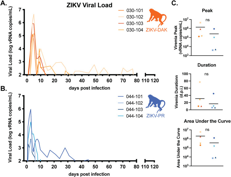
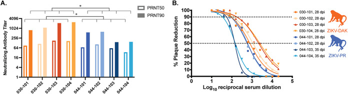
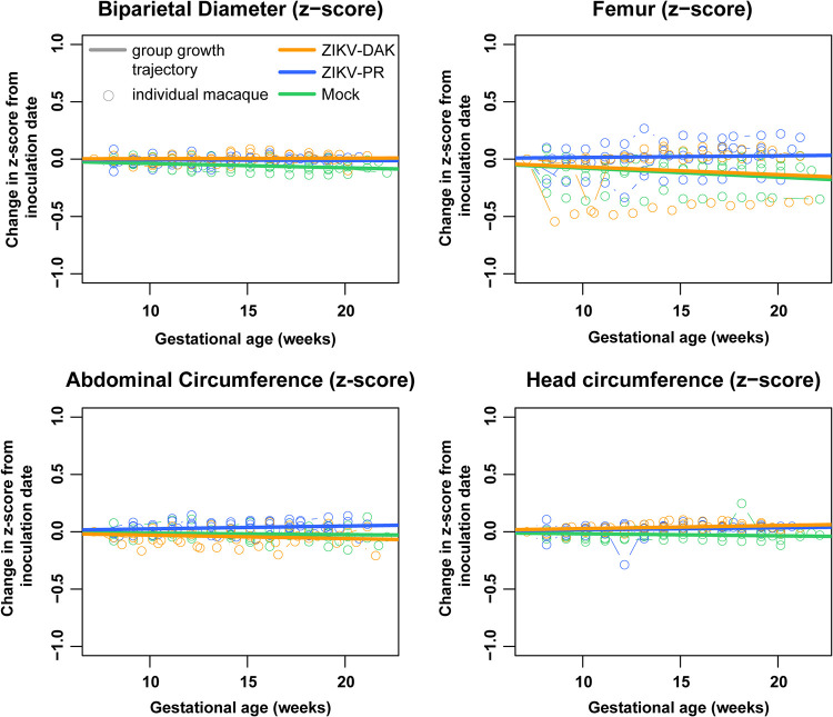
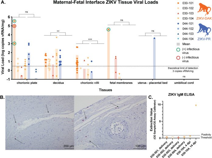
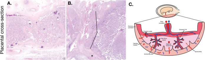
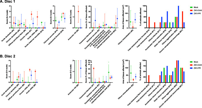
Following the Zika virus (ZIKV) outbreak in the Americas, ZIKV was causally associated with microcephaly and a range of neurological and developmental symptoms, termed congenital Zika syndrome (CZS). The viruses responsible for this outbreak belonged to the Asian lineage of ZIKV. However, in vitro and in vivo studies assessing the pathogenesis of African-lineage ZIKV demonstrated that African-lineage isolates often replicated to high titers and caused more-severe pathology than Asian-lineage isolates. To date, the pathogenesis of African-lineage ZIKV in a translational model, particularly during pregnancy, has not been rigorously characterized. Here, we infected four pregnant rhesus macaques with a low-passage-number strain of African-lineage ZIKV and compared its pathogenesis to those for a cohort of four pregnant rhesus macaques infected with an Asian-lineage isolate and a cohort of mock-inoculated controls. The viral replication kinetics for the two experimental groups were not significantly different, and both groups developed robust neutralizing antibody titers above levels considered to be protective. There was no evidence of significant fetal head growth restriction or gross fetal harm at delivery (1 to 1.5 weeks prior to full term) in either group. However, a significantly higher burden of ZIKV viral RNA (vRNA) was found in the maternal-fetal interface tissues of the macaques exposed to an African-lineage isolate. Our findings suggest that ZIKV of any genetic lineage poses a threat to pregnant individuals and their infants. IMPORTANCE ZIKV was first identified in 1947 in Africa, but most of our knowledge of ZIKV is based on studies of the distinct Asian genetic lineage, which caused the outbreak in the Americas in 2015 to 2016. In its most recent update, the WHO stated that improved understanding of African-lineage ZIKV pathogenesis during pregnancy must be a priority. The recent detection of African-lineage isolates in Brazil underscores the need to understand the impact of these viruses. Here, we provide the first comprehensive assessment of African-lineage ZIKV infection during pregnancy in a translational nonhuman primate model. We show that African-lineage isolates replicate with kinetics similar to those of Asian-lineage isolates and can infect the placenta. However, there was no evidence of more-severe outcomes with African-lineage isolates. Our results highlight both the threat that African-lineage ZIKV poses to pregnant individuals and their infants and the need for epidemiological and translational in vivo studies with African-lineage ZIKV.

Keywords: ZIKV; Zika virus; arbovirus; congenital Zika syndrome; flavivirus; macaque; pregnant.


Figures

FIG 1
FIG 1
Study overview. Groups of four pregnant macaques were challenged between gestational days 45 and 50 (late first trimester) with either ZIKV-DAK, ZIKV-PR, or PBS (mock). Following viral challenge, blood was collected daily from 0 to 10 dpi, then twice weekly until viremia resolved, and then once weekly until delivery. Ultrasound was performed once weekly to measure fetal health and growth. Between gestational days 155 and 160 (1 to 1.5 weeks prior to full term), infants were delivered via cesarean section (C-section), and maternal-fetal interface tissues, including the placenta, fetal membranes, umbilical cord, and placental bed, were collected. Infants born to dams inoculated with ZIKV-DAK were humanely euthanized, and a comprehensive set of tissues was collected. Infants born to dams challenged with ZIKV-PR or PBS (mock) were paired with their mothers and followed for long-term behavioral analysis. Data from the long-term behavioral analysis will be published as part of a separate study.
FIG 2
FIG 2
Replication kinetics of ZIKV-DAK and ZIKV-PR. (A and B) Viral loads were determined using ZIKV-specific QRT-PCR of RNA isolated from plasma. Only values above the assay’s limit of quantification (100 copies/ml) are shown. (C) There were no statistically significant differences in the peak, duration, or area under the curve of viremia between the two groups (by two-sample t tests).
FIG 3
FIG 3
Neutralizing antibody titers. (A) Plaque reduction neutralization tests (PRNT) were performed on serum samples collected between 28 and 35 days postinfection to determine the titers of ZIKV-specific neutralizing antibodies. (B) Neutralization curves were constructed using GraphPad Prism. PRNT90 and PRNT50 values were estimated using nonlinear regression analysis and are shown on a bar graph (A) and indicated with dotted lines (B). PRNT50 and PRNT90 were compared using an unpaired parametric t test. ZIKV-DAK-infected macaques had significantly higher PRNT50 (P = 0.0371) and PRNT90 (P = 0.0243) than ZIVK-PR-infected macaques.
FIG 4
FIG 4
Intrauterine fetal growth. Sonographic imaging was performed weekly to measure fetal health and growth. Normative measurement data from the California National Primate Research Center were used to calculate Z-scores for each weekly measurement for each macaque. The change in the Z-score from the baseline measurement is plotted for each macaque with an open circle. Growth trajectories were quantified by calculating the regression slope parameters from baseline for each experimental group (solid lines) using a linear mixed-effects model with animal-specific random effects and an autoregressive correlation structure. Compared to the normative data, mock-inoculated animals had significantly reduced biparietal diameter growth (P = 0.0207); ZIKV-PR- and ZIKV-DAK-inoculated animals had very modest, but statistically significant, increases in head circumference growth (P, 0.0230 and 0.0179, respectively).
FIG 5
FIG 5
vRNA at the maternal-fetal interface. For each macaque, tissue biopsy specimens were collected from the chorionic plate, chorionic villi, and decidua from each placental cotyledon; one to three biopsy specimens were collected from the fetal membranes; and one biopsy specimen was collected from the uterine placental bed and umbilical cord. Viral loads were determined by ZIKV-specific QRT-PCR from RNA isolated from tissue samples. (A) Viral loads of maternal-fetal interface tissues. A nonparametric Mann-Whitney test was used to assess the statistical significance of differences between the experimental groups in samples containing more than the theoretical limit of detection of 3 copies vRNA/mg tissue (**, P < 0.01, ***, P < 0.001; ns, not significant). (B) Representative images from in situ hybridization performed on fixed tissue sections from each of the placental cotyledons from macaque 030-101. Positive staining for ZIKV RNA (red; indicated by arrows) was identified in 11 of the 17 cotyledons tested, primarily in the chorionic plate. (C) Serum samples from the four infants born to ZIKV-DAK-infected mothers were tested via ELISA for the presence of ZIKV IgM antibodies. Serum samples from dam 030-104 were included as positive (14 dpi) and negative (4 dpi) controls.
FIG 6
FIG 6
Representative images of ZIKV-DAK placental pathology. (A) Normal placental cross section. The decidua basalis is located at the bottom of the cross section; the chorionic plate is located at the top. The placental parenchyma has a spongy appearance as a result of a robust network of villi. (B) Transmural infarction was noted in 4 of 4 macaques infected with ZIKV-DAK. Lines indicate the region of infarcted tissue (area of nonfunctional, ischemic, villous parenchyma), and the asterisk indicates the trophoblastic shell of the basal plate. (C) Graphical representation of a cross section of the placenta. The orientation of the diagram corresponds to the orientation of the cross sections in panels A and B.
FIG 7
FIG 7
Placental pathology scoring. The central cross section of each placental disc was evaluated for 22 pathological features. Some features were specific to the fetal membranes or uterus; these are found only under the disc 1 scoring. Statistical pairwise comparisons between groups were performed for each feature. For quantitative features (features 1 to 9, 12 to 16, and 22), a nonparametric Wilcoxon rank sum test was used; for binary features (features 10, 11, and 17 to 21), Fisher’s exact test was used. For quantitative features, the median value is shown, with error bars representing the interquartile range. Pairwise comparisons revealed no statistically significant differences for any of the features.

Similar articles

Cited by

  • Fetal loss in pregnant rhesus macaques infected with high-dose African-lineage Zika virus.
    Raasch LE, Yamamoto K, Newman CM, Rosinski JR, Shepherd PM, Razo E, Crooks CM, Bliss MI, Breitbach ME, Sneed EL, Weiler AM, Zeng X, Noguchi KK, Morgan TK, Fuhler NA, Bohm EK, Alberts AJ, Havlicek SJ, Kabakov S, Mitzey AM, Antony KM, Ausderau KK, Mejia A, Basu P, Simmons HA, Eickhoff JC, Aliota MT, Mohr EL, Friedrich TC, Golos TG, O'Connor DH, Dudley DM. Raasch LE, et al. PLoS Negl Trop Dis. 2022 Aug 4;16(8):e0010623. doi: 10.1371/journal.pntd.0010623. eCollection 2022 Aug. PLoS Negl Trop Dis. 2022. PMID: 35926066 Free PMC article.
  • Human immune globulin treatment controls Zika viremia in pregnant rhesus macaques.
    Dudley DM, Koenig MR, Stewart LM, Semler MR, Newman CM, Shepherd PM, Yamamoto K, Breitbach ME, Schotzko M, Kohn S, Antony KM, Qiu H, Tunga P, Anderson DM, Guo W, Dennis M, Singh T, Rybarczyk S, Weiler AM, Razo E, Mitzey A, Zeng X, Eickhoff JC, Mohr EL, Simmons HA, Fritsch MK, Mejia A, Aliota MT, Friedrich TC, Golos TG, Kodihalli S, Permar SR, O'Connor DH. Dudley DM, et al. PLoS One. 2022 Jul 14;17(7):e0266664. doi: 10.1371/journal.pone.0266664. eCollection 2022. PLoS One. 2022. PMID: 35834540 Free PMC article.
  • Control of maternal Zika virus infection during pregnancy is associated with lower antibody titers in a macaque model.
    Krabbe NP, Razo E, Abraham HJ, Spanton RV, Shi Y, Bhattacharya S, Bohm EK, Pritchard JC, Weiler AM, Mitzey AM, Eickhoff JC, Sullivan E, Tan JC, Aliota MT, Friedrich TC, O'Connor DH, Golos TG, Mohr EL. Krabbe NP, et al. Front Immunol. 2023 Sep 22;14:1267638. doi: 10.3389/fimmu.2023.1267638. eCollection 2023. Front Immunol. 2023. PMID: 37809089 Free PMC article.
  • Frequent first-trimester pregnancy loss in rhesus macaques infected with African-lineage Zika virus.
    Rosinski JR, Raasch LE, Barros Tiburcio P, Breitbach ME, Shepherd PM, Yamamoto K, Razo E, Krabbe NP, Bliss MI, Richardson AD, Einwalter MA, Weiler AM, Sneed EL, Fuchs KB, Zeng X, Noguchi KK, Morgan TK, Alberts AJ, Antony KM, Kabakov S, Ausderau KK, Bohm EK, Pritchard JC, Spanton RV, Ver Hoove JN, Kim CBY, Nork TM, Katz AW, Rasmussen CA, Hartman A, Mejia A, Basu P, Simmons HA, Eickhoff JC, Friedrich TC, Aliota MT, Mohr EL, Dudley DM, O'Connor DH, Newman CM. Rosinski JR, et al. PLoS Pathog. 2023 Mar 28;19(3):e1011282. doi: 10.1371/journal.ppat.1011282. eCollection 2023 Mar. PLoS Pathog. 2023. PMID: 36976812 Free PMC article.
  • Early Embryonic Loss Following Intravaginal Zika Virus Challenge in Rhesus Macaques.
    Newman CM, Tarantal AF, Martinez ML, Simmons HA, Morgan TK, Zeng X, Rosinski JR, Bliss MI, Bohm EK, Dudley DM, Aliota MT, Friedrich TC, Miller CJ, O'Connor DH. Newman CM, et al. Front Immunol. 2021 May 17;12:686437. doi: 10.3389/fimmu.2021.686437. eCollection 2021. Front Immunol. 2021. PMID: 34079560 Free PMC article.

References

    1. Rasmussen SA, Jamieson DJ, Honein MA, Petersen LR. 2016. Zika virus and birth defects—reviewing the evidence for causality. N Engl J Med 374:1981–1987. 10.1056/NEJMsr1604338. - DOI - PubMed
    1. Musso D, Ko AI, Baud D. 2019. Zika virus infection—after the pandemic. N Engl J Med 381:1444–1457. 10.1056/NEJMra1808246. - DOI - PubMed
    1. Grubaugh ND, Saraf S, Gangavarapu K, Watts A, Tan AL, Oidtman RJ, Ladner JT, Oliveira G, Matteson NL, Kraemer MUG, Vogels CBF, Hentoff A, Bhatia D, Stanek D, Scott B, Landis V, Stryker I, Cone MR, Kopp EW, Cannons AC, Heberlein-Larson L, White S, Gillis LD, Ricciardi MJ, Kwal J, Lichtenberger PK, Magnani DM, Watkins DI, Palacios G, Hamer DH, GeoSentinel Surveillance Network, Gardner LM, Perkins TA, Baele G, Khan K, Morrison A, Isern S, Michael SF, Andersen KG. 2019. Travel surveillance and genomics uncover a hidden Zika outbreak during the waning epidemic. Cell 178:1057–1071.e11. 10.1016/j.cell.2019.07.018. - DOI - PMC - PubMed
    1. Yuan L, Huang XY, Liu ZY, Zhang F, Zhu XL, Yu JY, Ji X, Xu YP, Li G, Li C, Wang HJ, Deng YQ, Wu M, Cheng ML, Ye Q, Xie DY, Li XF, Wang X, Shi W, Hu B, Shi PY, Xu Z, Qin CF. 2017. A single mutation in the prM protein of Zika virus contributes to fetal microcephaly. Science 358:933–936. 10.1126/science.aam7120. - DOI - PubMed
    1. Simonin Y, Loustalot F, Desmetz C, Foulongne V, Constant O, Fournier-Wirth C, Leon F, Molès JP, Goubaud A, Lemaitre JM, Maquart M, Leparc-Goffart I, Briant L, Nagot N, Van de Perre P, Salinas S. 2016. Zika virus strains potentially display different infectious profiles in human neural cells. EBioMedicine 12:161–169. 10.1016/j.ebiom.2016.09.020. - DOI - PMC - PubMed

Publication types