. 2021 Jun 16;34(3):e0018820.
doi: 10.1128/CMR.00188-20. Epub 2021 Jun 2.

Mobile Oxazolidinone Resistance Genes in Gram-Positive and Gram-Negative Bacteria

Affiliations

Mobile Oxazolidinone Resistance Genes in Gram-Positive and Gram-Negative Bacteria

Stefan Schwarz et al. Clin Microbiol Rev. .

Abstract

Seven mobile oxazolidinone resistance genes, including cfr, cfr(B), cfr(C), cfr(D), cfr(E), optrA, and poxtA, have been identified to date. The cfr genes code for 23S rRNA methylases, which confer a multiresistance phenotype that includes resistance to phenicols, lincosamides, oxazolidinones, pleuromutilins, and streptogramin A compounds. The optrA and poxtA genes code for ABC-F proteins that protect the bacterial ribosomes from the inhibitory effects of oxazolidinones. The optrA gene confers resistance to oxazolidinones and phenicols, while the poxtA gene confers elevated MICs or resistance to oxazolidinones, phenicols, and tetracycline. These oxazolidinone resistance genes are most frequently found on plasmids, but they are also located on transposons, integrative and conjugative elements (ICEs), genomic islands, and prophages. In these mobile genetic elements (MGEs), insertion sequences (IS) most often flanked the cfr, optrA, and poxtA genes and were able to generate translocatable units (TUs) that comprise the oxazolidinone resistance genes and occasionally also other genes. MGEs and TUs play an important role in the dissemination of oxazolidinone resistance genes across strain, species, and genus boundaries. Most frequently, these MGEs also harbor genes that mediate resistance not only to antimicrobial agents of other classes, but also to metals and biocides. Direct selection pressure by the use of antimicrobial agents to which the oxazolidinone resistance genes confer resistance, but also indirect selection pressure by the use of antimicrobial agents, metals, or biocides (the respective resistance genes against which are colocated on cfr-, optrA-, or poxtA-carrying MGEs) may play a role in the coselection and persistence of oxazolidinone resistance genes.

Keywords: cfr; genomic island; horizontal transfer; integrative and conjugative element; mobile genetic element; optrA; oxazolidinones; plasmid; poxtA; prophage; transposon.


Figures

FIG 1
FIG 1
Schematic presentation of the mode of action of oxazolidinones. Oxazolidinones inhibit protein biosynthesis by preventing the formation of a functional 70S initiation complex composed of the 30S ribosomal subunit, initiation factors, mRNA, formylmethionyl-tRNA (fMET-tRNA), and the 50S ribosomal subunit. (Based on data from reference .)
FIG 2
FIG 2
Percentages of linezolid-susceptible S. aureus, coagulase-negative staphyloccoci (CoNS), and Enterococcus isolates as determined in the worldwide Zyvox Annual Appraisal of Potency and Spectrum (ZAAPS) program during the years 2002 to 2016 (58–66, 69–71).
FIG 3
FIG 3
Geographical distribution of cfr-carrying bacteria. The countries in blue are those from which the occurrence of cfr-carrying bacteria has been reported.
FIG 4
FIG 4
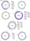
Structural comparison of cfr-carrying staphylococcal plasmids (constructed by BLAST Ring Image Generator [BRIG]). Relevant genes with known functions and insertion elements are indicated for the respective reference plasmid in the outer ring. The cfr and the optrA genes are indicated in red. The innermost circle provides a size scale, while the next innermost circle shows the GC content. Other plasmids used for comparison (if available) are indicated by color-coded rings, with the reference plasmid representing the innermost colored ring. (A) pSAM13-0451, pSS-03 (Staphylococcus arlettae), pSS-03 (Staphylococcus cohnii), and pMSA16, (B) pERGB, (C) pSCFS1, (D) pSAM12-0145, pSA737, pSEPI8573, p14-01514, pY96A, p12-02300, and unnamed_1, (E) pH29-46, pk8D6P-cfr, pY8P168P-cfr, pSX01, pLRSA417, pSR01, and p12-00322, (F) and pWo28-1, pWo28-3, and pWo27-9, as well as (G) pSA-01.
FIG 5
FIG 5
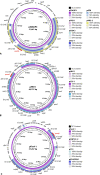
Structural comparison of cfr-carrying enterococcal plasmids (constructed by BRIG). Relevant genes with known functions and insertion elements are indicated for the respective reference plasmid in the outer ring. The cfr gene is indicated in red. The innermost circle provides a size scale, while the next innermost circle shows the GC content. Other plasmids used for comparison (if available) are indicated by color-coded rings, with the reference plasmid representing the innermost colored ring. (A) pEF-01, pCPPF5, pE30, and unnamed, (B) p4, pF120805 and pE35048-oc, (C) pFSIS1608820, and (D) pFas4-2.
FIG 6
FIG 6
Structural comparison of cfr-carrying plasmids in E. coli and Proteus spp. (constructed by BRIG). Relevant genes with known functions and insertion elements are indicated for the respective reference plasmid in the outer ring. The cfr gene is indicated in red. The innermost circle provides a size scale, while the next innermost circle shows the GC content. Other plasmids used for comparison (if available) are indicated by color-coded rings, with the reference plasmid representing the innermost colored ring. (A) pGXEC3, pGXEC6, pSD11, pEC14cfr, and pHNEP129 (all E. coli), (B) pHNFP671, pHNEP124, and pFSEC-01 (all E. coli), (C) pSCEC2 (E. coli), and (D) pZF1-cfr, pZF2-cfr, pGE32-51, and p52 (all P. cibarius).
FIG 7
FIG 7
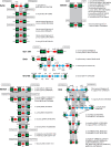
Insertion sequences flanking the cfr gene. The insertion sequences IS256, ISEnfa5, IS21-558, IS431, IS1216E, ISEnfa4, and IS26 are displayed as black boxes, with the green arrow(s) inside symbolizing the respective transposase gene(s). The cfr gene is shown as a red arrow. Additional genes are shown as blue arrows. In all cases, the arrowhead indicates the direction of transcription. Whenever direct repeats were identified at the termini of the IS elements that flank the cfr region, they are indicated in boxes. The gray-shaded area indicates >99% nucleotide sequence identity. For each specific IS-cfr-IS arrangement, the bacterial species, the location (plasmid/chromosomal DNA) and the database accession number (in brackets) are given on the righthand side.
FIG 8
FIG 8
Structural comparison of cfr(C)-carrying plasmids in C. coli (constructed by BRIG). Relevant genes with known functions and insertion elements are indicated for the respective reference plasmid in the outer ring. The cfr(C) gene is indicated in red. The innermost circle provides a size scale, while the next innermost circle shows the GC content. Other plasmids used for comparison are indicated by color-coded rings, with the reference plasmid representing the innermost colored ring. The plasmids used for this comparison are pN46788F, pN61740F, pN61925F, and pTx-40.
FIG 9
FIG 9
Geographical distribution of optrA-carrying bacteria. The countries in blue are those from which the occurrence of optrA-carrying bacteria has been reported.
FIG 10
FIG 10
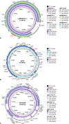
Structural comparison of optrA-carrying plasmids in enterococci (constructed by BRIG). Relevant genes with known functions and insertion elements are indicated for the respective reference plasmid in the outer ring. The optrA gene is indicated in red. The innermost circle provides a size scale, while the next innermost circle shows the GC content. Other plasmids used for comparison are indicated by color-coded rings, with the reference plasmid representing the innermost colored ring. (A) pN60443F-2, pN48037F-3, pEFs17-1, p6742_1, pM17/0149, pKUB3006-4, and pKUB3007-4, and (B) pC54 and pC25-1, as well as (C) unnamed, pBA17124_P1, pBP5067_P1, pM17/0314, and p15-307-1_02.
FIG 11
FIG 11
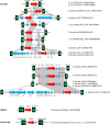
Insertion sequences flanking the optrA gene. The insertion sequences IS1216E, ISEfa15, and ISChh1-like are displayed as black boxes with the green arrow(s) inside symbolizing the respective transposase gene(s). The optrA gene is shown as a red arrow. Additional resistance genes, such as fexA and erm(A)-like, are displayed as rose arrows, while other genes are shown as blue arrows. In all cases, the arrowhead indicates the direction of transcription. Whenever direct repeats were identified at the termini of the IS elements that flank the cfr region, they are indicated in boxes. The gray-shaded area indicates >99% nucleotide sequence identity. For each specific IS-cfr-IS arrangement, the bacterial species, the location (plasmid/integrative and conjugative element [ICE]/chromosomal multidrug resistance genomic island [MDRGI]) and the database accession number (in brackets) are given on the righthand side.
FIG 12
FIG 12
Geographical distribution of poxtA-carrying bacteria. The countries in blue are those from which the occurrence of poxtA-carrying bacteria has been reported.
FIG 13
FIG 13
Structural comparison of poxtA-carrying plasmids in enterococci (constructed by BRIG). Relevant genes with known functions and insertion elements are indicated for the respective reference plasmid in the outer ring. The poxtA gene is indicated in red. The innermost circle provides a size scale, while the next innermost circle shows the GC content. Other plasmids used for comparison are indicated by color-coded rings, with the reference plasmid representing the innermost colored ring. (A) pSDGJP3, pYN2-1, pSCBC1, pSC3-1, pGZ8, and pHB2-2, and (B) pHN11, pC25-1, and pC27-2, as well as (C) pFas4-2, pCQP3-9_2, pC10, and pM16/0594.
FIG 14
FIG 14
Insertion sequences flanking the poxtA gene. The insertion sequences IS1216E and IS1252 are displayed as black boxes, with the green arrow inside symbolizing the respective transposase gene. The poxtA gene is shown as a red arrow. The additional resistance gene fexB is displayed as a rose arrow, while other genes are shown as blue arrows. In all cases, the arrowhead indicates the direction of transcription. The gray-shaded area indicates >99% nucleotide sequence identity. For each specific IS-cfr-IS arrangement, the bacterial species, the plasmid on which it is located, and the database accession number (in brackets) are given on the righthand side.

Similar articles

Cited by

References

    1. Slee AM, Wuonola MA, McRipley RJ, Zajac I, Zawada MJ, Bartholomew PT, Gregory WA, Forbes M. 1987. Oxazolidinones, a new class of synthetic antibacterial agents: in vitro and in vivo activities of DuP 105 and DuP 721. Antimicrob Agents Chemother 31:1791–1797. 10.1128/aac.31.11.1791. - DOI - PMC - PubMed
    1. Brickner SJ, Hutchinson DK, Barbachyn MR, Manninen PR, Ulanowicz DA, Garmon SA, Grega KC, Hendges SK, Toops DS, Ford CW, Zurenko GE. 1996. Synthesis and antibacterial activity of U-100592 and U-100766, two oxazolidinone antibacterial agents for the potential treatment of multidrug-resistant Gram-positive bacterial infections. J Med Chem 39:673–679. 10.1021/jm9509556. - DOI - PubMed
    1. Zurenko GE, Yagi BH, Schaadt RD, Allison JW, Kilburn JO, Glickman SE, Hutchinson DK, Barbachyn MR, Brickner SJ. 1996. In vitro activities of U-100592 and U-100766, novel oxazolidinone antibacterial agents. Antimicrob Agents Chemother 40:839–845. 10.1128/AAC.40.4.839. - DOI - PMC - PubMed
    1. Bain KT, Wittbrodt ET. 2001. Linezolid for the treatment of resistant Gram-positive cocci. Ann Pharmacother 35:566–575. 10.1345/aph.10276. - DOI - PubMed
    1. Tsuji BT, Kaatz GW, Ryback MJ. 1999. –2021. Linezolid and other oxazolidinones. In Antimicrobial therapy and vaccines volume II: antimicrobial agents. Antimicrobe, Pittsburgh, PA. http://www.antimicrobe.org/d13.asp. Accessed 12 Jan 2021.

Publication types

MeSH terms

LinkOut - more resources