Review
. 2021 Aug 18;85(3):e0006421.
doi: 10.1128/MMBR.00064-21. Epub 2021 Jun 2.

Large Clostridial Toxins: Mechanisms and Roles in Disease

Affiliations
Review

Large Clostridial Toxins: Mechanisms and Roles in Disease

Kathleen E Orrell et al. Microbiol Mol Biol Rev. .

Abstract

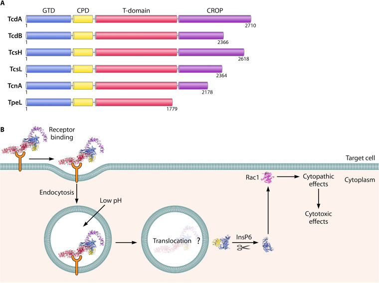
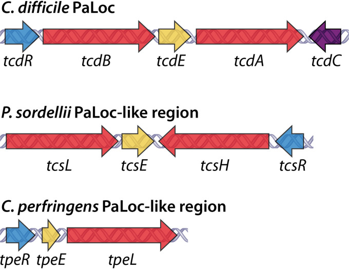
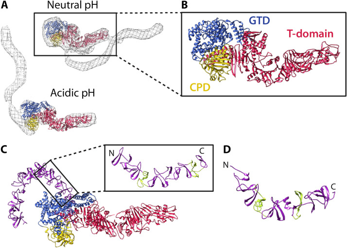
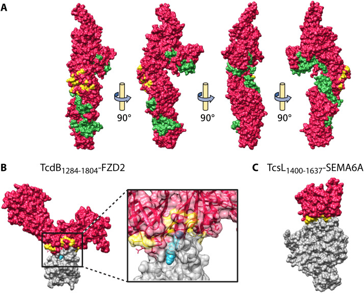
Large clostridial toxins (LCTs) are a family of bacterial exotoxins that infiltrate and destroy target cells. Members of the LCT family include Clostridioides difficile toxins TcdA and TcdB, Paeniclostridium sordellii toxins TcsL and TcsH, Clostridium novyi toxin TcnA, and Clostridium perfringens toxin TpeL. Since the 19th century, LCT-secreting bacteria have been isolated from the blood, organs, and wounds of diseased individuals, and LCTs have been implicated as the primary virulence factors in a variety of infections, including C. difficile infection and some cases of wound-associated gas gangrene. Clostridia express and secrete LCTs in response to various physiological signals. LCTs invade host cells by binding specific cell surface receptors, ultimately leading to internalization into acidified vesicles. Acidic pH promotes conformational changes within LCTs, which culminates in translocation of the N-terminal glycosyltransferase and cysteine protease domain across the endosomal membrane and into the cytosol, leading first to cytopathic effects and later to cytotoxic effects. The focus of this review is on the role of LCTs in infection and disease, the mechanism of LCT intoxication, with emphasis on recent structural work and toxin subtyping analysis, and the genomic discovery and characterization of LCT homologues. We provide a comprehensive review of these topics and offer our perspective on emerging questions and future research directions for this enigmatic family of toxins.

Keywords: Clostridium difficile; large clostridial toxin; toxin; toxin-mediated diseases; toxin-receptor interaction.


Figures

FIG 1
FIG 1
LCT domain architecture and mechanism of action. (A) Individual domains are colored as follows: glycosyltransferase domain (GTD) in blue, cysteine protease domain (CPD) in yellow, translocation and receptor-binding domain (T domain) in red, and combined repeating oligopeptide (CROP) in purple. (B) Schematic illustrating the major steps of LCT intoxication of host cells, using TcdB (PDB 6OQ5).
FIG 2
FIG 2
LCT gene organization. The PaLoc of C. difficile and PaLoc-like regions of P. sordellii and C. perfringens, first identified in C. difficile strain VPI 10463, P. sordellii strain VPI 9048, and C. perfringens strain ATCC 3626. LCT genes are colored red. Accessory genes are colored as follows: alternative σ factor (tcdR/tcsR/tpeR) in blue, anti-σ factor (tcdC) in purple, and holin-like protein (tcdE/tcsE/tpeE) in yellow.
FIG 3
FIG 3
Structure and organization of the C. difficile holotoxin. (A) TcdA1–1832 (PDB 4R04) fit into the EM map of the TcdA holotoxin at neutral and acidic pH (140) using Chimera (267). The TcdA EM maps were kindly provided by Borden Lacy and are reproduced with permission. (B) Focused view of TcdA1–1832. (C) Structure of TcdB1–2366 at acidic pH (PDB 6OQ5). In the focused image, short repeats (SRs) are colored purple and long repeats (LRs) are colored green. (D) Structure of a fragment of the TcdA CROP (PDB 2G7C), with SRs colored purple and LRs colored green.
FIG 4
FIG 4
Structure of the LCT T domain and LCT-receptor complexes. (A) T domain of TcdB (PDB 6OQ5), with the hydrophobic helical stretch (residues 956 to 1135) colored green and the interface residues for FZD2 binding colored yellow. (B) TcdB1284–1804-FZD2 (PDB 6C0B), with focus on the receptor-binding interface. TcdB is colored red, FZD2 interface residues on TcdB are colored yellow, FZD2 is colored gray, and palmitoleic acid (PAM) is colored blue. (C) TcsL1400–1637-SEMA6A (PDB 6WTS). TcsL is colored red, SEMA6A interface residues on TcsL are colored yellow, and SEMA6A is colored gray.
FIG 5
FIG 5
Translocation features of the TcdA T domain. (A) Structure of the TcdA T domain (PDB 4R04), with the evolutionarily conserved translocase colored purple (residues 853 to 1475) and the hydrophobic region (residues 958 to 1137) colored green. (B) Focus on the evolutionarily conserved region and (C) the hydrophobic region without the rest of the T domain. Residues with important functions in translocation identified by Zhang et al. (197) are shown as pink sticks.
FIG 6
FIG 6
Structural features of the C. difficile toxin cysteine protease domain. (A) TcdA cysteine protease domain (PDB 3HO6) and (B) TcdB cysteine protease domain (PDB 3PEE) bound to InsP6 (268). The β-flap is colored purple, residues of the catalytic triad are depicted as blue sticks, and InsP6 is colored green.
FIG 7
FIG 7
Structural features of the LCT glycosyltransferase domain. (A) TcdB glycosyltransferase domain (PDB 2BVM) in complex with the cosubstrate and a manganese ion, depicted as green sticks and a sphere, respectively. Residues important for catalytic function are depicted as yellow sticks. Structures of the (B) TcdA (PDB 3SRZ), (C) TcsL (PDB 2VKD), and (D) TcnA (PDB 2VK9) glycosyltransferase domains. The membrane localization domain (MLD) is colored aqua.

References

    1. Savva CG, Clark AR, Naylor CE, Popoff MR, Moss DS, Basak AK, Titball RW, Bokori-Brown M. 2019. The pore structure of Clostridium perfringens epsilon toxin. Nat Commun 10:2641. 10.1038/s41467-019-10645-8. - DOI - PMC - PubMed
    1. Gerding DN, Johnson S, Rupnik M, Aktories K. 2014. Clostridium difficile binary toxin CDT: mechanism, epidemiology, and potential clinical importance. Gut Microbes 5:15–27. 10.4161/gmic.26854. - DOI - PMC - PubMed
    1. Bette P, Oksche A, Mauler F, Eichel-Streiber Cv, Popoff MR, Habermann E. 1991. A comparative biochemical, pharmacological and immunological study of Clostridium novyi alpha-toxin, C. difficile toxin B and C. sordellii lethal toxin. Toxicon 29:877–887. 10.1016/0041-0101(91)90224-F. - DOI - PubMed
    1. Aronoff DM, Kazanjian PH. 2018. Historical and contemporary features of infections due to Clostridium novyi. Anaerobe 50:80–84. 10.1016/j.anaerobe.2017.12.012. - DOI - PubMed
    1. Aldape MJ, Bryant AE, Stevens DL. 2006. Clostridium sordellii infection: epidemiology, clinical findings, and current perspectives on diagnosis and treatment. Clin Infect Dis 43:1436–1446. 10.1086/508866. - DOI - PubMed

MeSH terms

Grants and funding

LinkOut - more resources