Review
. 2022 Mar;387(3):319-336.
doi: 10.1007/s00441-021-03477-w. Epub 2021 Jun 2.

New insights into glial scar formation after spinal cord injury

Affiliations
Review

New insights into glial scar formation after spinal cord injury

Amanda Phuong Tran et al. Cell Tissue Res. 2022 Mar.

Abstract

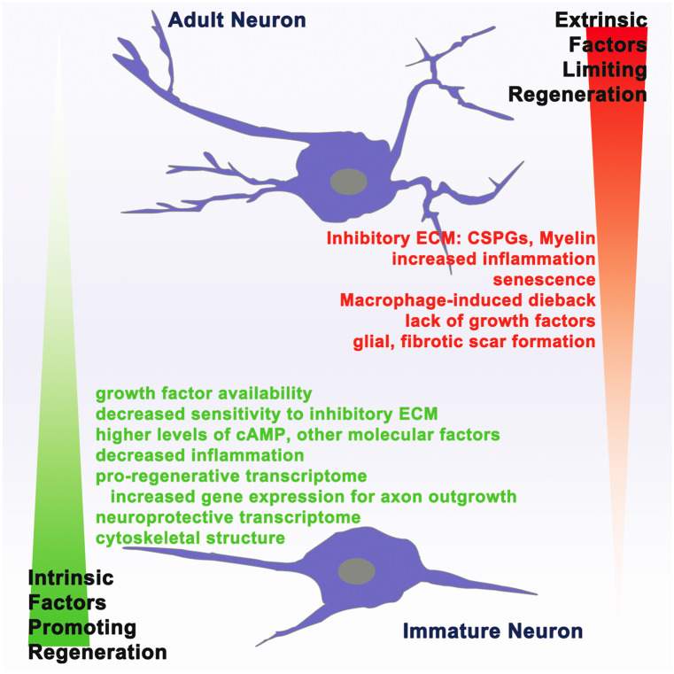
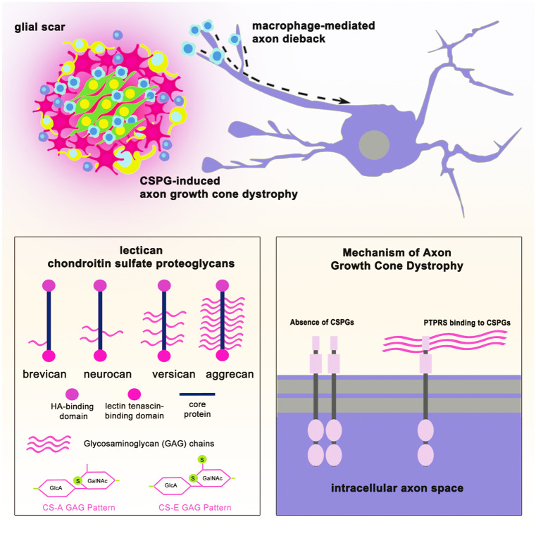
Severe spinal cord injury causes permanent loss of function and sensation throughout the body. The trauma causes a multifaceted torrent of pathophysiological processes which ultimately act to form a complex structure, permanently remodeling the cellular architecture and extracellular matrix. This structure is traditionally termed the glial/fibrotic scar. Similar cellular formations occur following stroke, infection, and neurodegenerative diseases of the central nervous system (CNS) signifying their fundamental importance to preservation of function. It is increasingly recognized that the scar performs multiple roles affecting recovery following traumatic injury. Innovative research into the properties of this structure is imperative to the development of treatment strategies to recover motor function and sensation following CNS trauma. In this review, we summarize how the regeneration potential of the CNS alters across phyla and age through formation of scar-like structures. We describe how new insights from next-generation sequencing technologies have yielded a more complex portrait of the molecular mechanisms governing the astrocyte, microglial, and neuronal responses to injury and development, especially of the glial component of the scar. Finally, we discuss possible combinatorial therapeutic approaches centering on scar modulation to restore function after severe CNS injury.

Keywords: Chondroitin sulfate proteoglycans; Glia; Glial scar; Glial scar formation; Single-cell RNA sequencing; Spinal cord injury.


Conflict of interest statement

A patent (9,937,242) filed by CWRU has been granted for ISP and licensed to NervGen Pharma Corp. The remaining authors declare no competing conflicts of interest.

Figures

Fig. 1
Fig. 1
Cellular constituents of the glial/fibrotic scar. Following spinal cord injury, pro-inflammatory cascades activate the multitude of cells found at the spinal cord lesion. The glial scar itself is composed of astrocytes, NG2 + oligodendrocyte progenitor cells, and microglia, among others. Astrocytes and oligodendrocyte progenitor cells of the lesion penumbra are especially important in remodeling the extracellular matrix and upregulating axon-inhibitory chondroitin sulfate proteoglycans (CSPGs). Cells of the lesion penumbra work in concert to cordon off the pro-inflammatory lesion epicenter. The lesion epicenter predominantly includes macrophages and fibroblasts which are sequestered to prevent the spread of inflammation after injury
Fig. 2
Fig. 2
Schematic phylogenetic relationship between regenerating animals. Simplified representation of the phylogenetic relationship between selected species capable of spinal cord regeneration (either within development or through their life span) following injury. Zebrafish, salamanders, and nemertea can regenerate their spinal cords throughout life following injury (red), frogs can do so at the tadpole stage (orange). Some higher-order animals are capable of this in the days following birth (green) including the opossum (until P17) and mouse (until P2). A number of other species (black) with common ancestors to these species are not known to regenerate their spinal cords following injury at any stage of development
Fig. 3
Fig. 3
Mammalian neurons lose regeneration potential with age. Embryonic and immature mammalian neurons have a higher potential for axon regeneration after injury due to increased pro-regenerative intrinsic factors and decreased regeneration-inhibitory extrinsic factors compared to the adult mammalian neuron. Immature neurons additionally possess pro-regenerative molecular signals/transcriptome, which is turned off as the neurons age into adulthood
Fig. 4
Fig. 4
Mechanisms of axon regeneration failure. Acutely after spinal cord injury, infiltrating macrophages come into physical contact with the tips of sheared axons causing the injured axon to dieback to the neuron soma. Chronically, chondroitin sulfate proteoglycans (CSPGs) of the glial scar cause growth cone dystrophy of approaching axons. CSPGs consist of a lectican group (brevican, neurocan, versican, and aggrecan) and phophocan and NG2 (not pictured here). The glycosaminoglycan (GAG) chains, notably sulfation patterns CS-A and CS-E, are especially inhibitory to axon regeneration. GAG chains of CSPGs bind to protein tyrosine phosphatase receptor sigma (PTPRS) promoting monomerization of the receptor at the axon growth cone to cause dystrophy and chronic regeneration failure

Similar articles

Cited by

References

    1. Alilain W. Shedding light on restoring respiratory function after spinal cord injury. Front Mol Neurosci. 2009;2:1–6. - PMC - PubMed
    1. Alilain WJ, Horn KP, Hu H, Dick TE, Silver J. Functional regeneration of respiratory pathways after spinal cord injury. Nat. 2011;475:196–200. - PMC - PubMed
    1. Anderson MA, Burda JE, Ren Y, Ao Y, O’Shea TM, Kawaguchi R, Coppola G, Khakh BS, Deming TJ, Sofroniew MV. Astrocyte scar formation aids central nervous system axon regeneration. Nature. 2016;532:195–200. - PMC - PubMed
    1. Andrews EM, Richards RJ, Yin FQ, Viapiano MS, Jakeman LB. Alterations in chondroitin sulfate proteoglycan expression occur both at and far from the site of spinal contusion injury. Exp Neurol. 2012;235:174–187. - PMC - PubMed
    1. Andrews MR, Soleman S, Cheah M, Tumbarello DA, Mason MR, Moloney E, Verhaagen J, Bensadoun JC, Schneider B, Aebischer P, Fawcett JW (2016) Axonal localization of integrins in the CNS is neuronal type and age dependent. eNeuro 3 - PMC - PubMed