

COVID-19 disrupts spermatogenesis through the oxidative stress pathway following induction of apoptosis
- PMID: 34076792
- PMCID: PMC8170653
- DOI: 10.1007/s10495-021-01680-2
COVID-19 disrupts spermatogenesis through the oxidative stress pathway following induction of apoptosis
Abstract
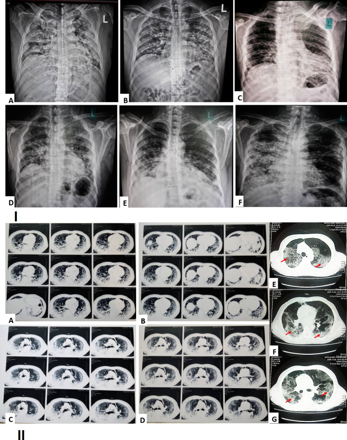
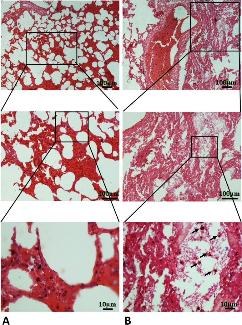
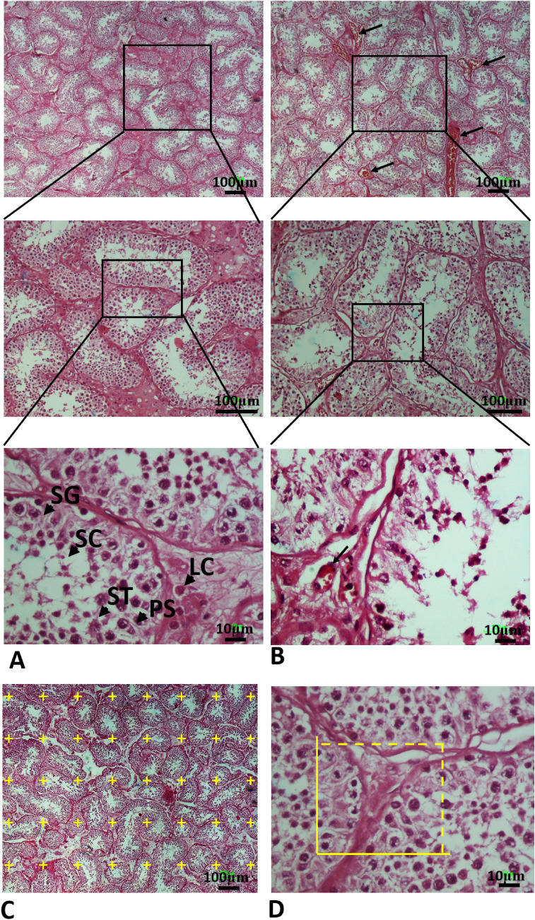
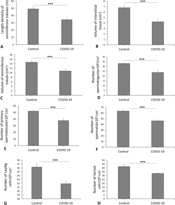
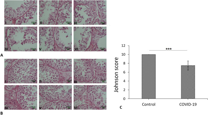
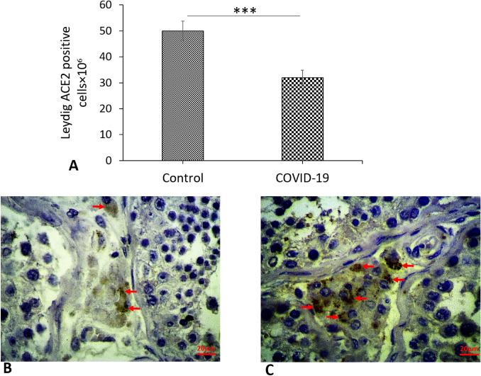
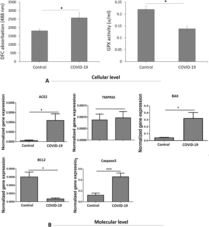
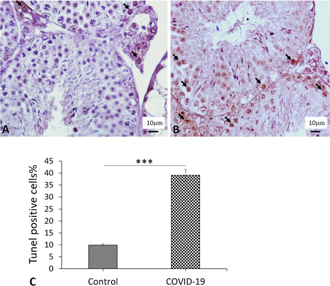
To evaluate the incidence of apoptosis within the testes of patients who died from severe acute respiratory syndrome coronavirus 2 (COVID-19) complications, testis tissue was collected from autopsies of COVID-19 positive (n = 6) and negative men (n = 6). They were then taken for histopathological experiments, and RNA extraction, to examine the expression of angiotensin-converting enzyme 2 (ACE2), transmembrane protease, serine 2 (TMPRSS2), BAX, BCL2 and Caspase3 genes. Reactive oxygen species (ROS) production and glutathione disulfide (GSH) activity were also thoroughly examined. Autopsied testicular specimens of COVID-19 showed that COVID-19 infection significantly decreased the seminiferous tubule length, interstitial tissue and seminiferous tubule volume, as well as the number of testicular cells. An analysis of the results showed that the Johnsen expressed a reduction in the COVID-19 group when compared to the control group. Our data showed that the expression of ACE2, BAX and Caspase3 were remarkably increased as well as a decrease in the expression of BCL2 in COVID-19 cases. Although, no significant difference was found for TMPRSS2. Furthermore, the results signified an increase in the formation of ROS and suppression of the GSH activity as oxidative stress biomarkers. The results of immunohistochemistry and TUNEL assay showed that the expression of ACE2 and the number of apoptotic cells significantly increased in the COVID-19 group. Overall, this study suggests that COVID-19 infection causes spermatogenesis disruption, probably through the oxidative stress pathway and subsequently induces apoptosis.
Keywords: Apoptosis; COVID-19; Spermatogenesis disrupt; Testicular tissue.
© 2021. The Author(s), under exclusive licence to Springer Science+Business Media, LLC, part of Springer Nature.
Conflict of interest statement
The authors declare that they have no competing interests.
Figures
Similar articles
-
Glutathione improves testicular spermatogenesis through inhibiting oxidative stress, mitochondrial damage, and apoptosis induced by copper deposition in mice with Wilson disease.Biomed Pharmacother. 2023 Feb;158:114107. doi: 10.1016/j.biopha.2022.114107. Epub 2022 Dec 8. Biomed Pharmacother. 2023. PMID: 36502753
-
In Silico Identification of miRNA-lncRNA Interactions in Male Reproductive Disorder Associated with COVID-19 Infection.Cells. 2021 Jun 12;10(6):1480. doi: 10.3390/cells10061480. Cells. 2021. PMID: 34204705 Free PMC article.
-
Nasopharyngeal Expression of Angiotensin-Converting Enzyme 2 and Transmembrane Serine Protease 2 in Children within SARS-CoV-2-Infected Family Clusters.Microbiol Spectr. 2021 Dec 22;9(3):e0078321. doi: 10.1128/Spectrum.00783-21. Epub 2021 Nov 3. Microbiol Spectr. 2021. PMID: 34730438 Free PMC article.
-
Perspectives into the possible effects of the B.1.1.7 variant of the severe acute respiratory syndrome coronavirus 2 (SARS-CoV-2) on spermatogenesis.J Basic Clin Physiol Pharmacol. 2021 Nov 26;33(1):9-12. doi: 10.1515/jbcpp-2021-0083. J Basic Clin Physiol Pharmacol. 2021. PMID: 34837491 Review.
-
Effects of COVID-19 on male sex function and its potential sexual transmission.Arch Ital Urol Androl. 2021 Mar 18;93(1):48-52. doi: 10.4081/aiua.2021.1.48. Arch Ital Urol Androl. 2021. PMID: 33754609
Cited by
-
COVID-19, oxidative stress, and male reproductive dysfunctions: is vitamin C a potential remedy?Physiol Res. 2022 Mar 25;71(1):47-54. doi: 10.33549/physiolres.934827. Epub 2022 Jan 19. Physiol Res. 2022. PMID: 35043653 Free PMC article. Review.
-
The role of oxidative stress in the pathogenesis of infections with coronaviruses.Front Microbiol. 2023 Jan 13;13:1111930. doi: 10.3389/fmicb.2022.1111930. eCollection 2022. Front Microbiol. 2023. PMID: 36713204 Free PMC article. Review.
-
Impact of COVID-19 on Male Fertility.Urology. 2022 Jun;164:33-39. doi: 10.1016/j.urology.2021.12.025. Epub 2022 Jan 8. Urology. 2022. PMID: 35007621 Free PMC article. Review.
-
Oxidative Stress Markers and Sperm DNA Fragmentation in Men Recovered from COVID-19.Int J Mol Sci. 2022 Sep 2;23(17):10060. doi: 10.3390/ijms231710060. Int J Mol Sci. 2022. PMID: 36077455 Free PMC article.
-
COVID-19 and Male Infertility: Is There a Role for Antioxidants?Antioxidants (Basel). 2023 Jul 25;12(8):1483. doi: 10.3390/antiox12081483. Antioxidants (Basel). 2023. PMID: 37627478 Free PMC article. Review.
References
-
- Organization WH. World Health Organization coronavirus disease 2019 (COVID-19) situation report. Geneva: World Health Organization; 2020.
MeSH terms
Substances
LinkOut - more resources
Full Text Sources
Medical
Research Materials
